
Forums › Laser Treatment Tips and Techniques › Soft Tissue Procedures › Treatment of Failing RCT
- This topic is empty.
-
AuthorPosts
-
Robert Gregg DDSSpectatorJeff,
Thanks for the post.
One of the frustrations I read on forums about the sinus tracts/fistulas is they won’t close up. Your post show how nicely these close with FRP Nd:YAG w/o burn or char, yet nice closure of the orifice.
What role do you think occlusal trauma is playing? Can you plint this tooth somehow?
Thanks for the post.
Bob
BenchwmerSpectatorBob,
The posterior teeth and bridge were extracted by the Periodontist (fractures, unrestorable) and treatment planned for implants. The patient didn’t want the grafts, etc. so what you see is what there is. Other than UR, she has an intact dentition.
All the anterior teeth are porcelain. She is happy with what is left. When something fails, then will be time for splints and PU. I’ve been waiting for over 5 years.
Jeff
BNelsonSpectatorJeff
Nice case. I did one just like it this week. Same setting s except I used 350 usec for hemostasis. Patient to come in next week for post op. Tooth is an abutment for a bridge so want to save it at all costs. I hope I have as good of luck as you had.
BenchwmerSpectatorBruce,
In some of these cases the infection will re-occur in 3-4 weeks. I re-treat with the same protocol, have had good luck with stable healing with no more than the 1 or 2 treatments.
Good luck.
Jeff
Lee AllenSpectatorJeff,
I am amazed at the results, but then, it is effective, I assume, because you reduced the bioburden.
What do you think was the original reason for the pa abscess, and will that need to be addressed for long term stability?
Would you expect similar results from any other type of laser?
Thanks for posting this interesting case.
BenchwmerSpectatorLee,
There is a breakdown somewhere in the patient host response. There seems to be a delicate balance between maintaining health and sliding to return of disease. Bob’s diagnosis of tramautic occlusion is probably dead-on. There are no Perio pockets. The treatment plan 7-8 years ago in this case was Perio Surgergy to save UR teeth, when they failed, the Periodontisr removed the failures. After extractions, implants were planned. When sinus lifts, etc were needed the patient decided to leave as is. Use of the PerioLase and LANAP techniques have maintained the rest of her dentition (sorry I didn’t have it 10 years ago).
The LANAP technique removes the diseased tissue, decontaminates the site and surrounding tissues and creates a closed system. This technique can only be done using a pulsed Nd:YAG with a short pulse width for ablation and a wide pulse width for hemostatis.
Since I am not changing the occlussion in this case, we will wait for the infection to return. We are at 3 months and counting. I’ll post the latest photos soon.
Jeff
Samuel MossSpectatorJeff,
Just my Ũ.02 worth. It appears that the original PA lesion had not healed and this is a failed root canal. How many have we seen over the years that were treated, the patient had no further symptomology, yet the lesion remained. I have seen some of mine and many of others over the years. There is no pain, no swelling, no tenderness. In those cases, why would you retreat the root canals, especially if the patient looks at you as if you had lost your mind? I had one last week where the patient had a root canal on #10 done many years ago. He had no swelling, no tenderness, no nothing. He fractured the crown and tooth off at the gumline. Strike 2 for the tooth. Told him that it was time for an implant since nothing left to cement to. He asked me to place a post and put him back together so that he could get his finances together for January to do the implant. I complied with his wishes. Two days later, he is swelling and hurting like a big dog. The root did not appear to be fractured–no verticle drop off probing, no movement. After antibiotics took hold, he came back in and was just tender at the apex of #10. I guess I just rattled it enough to set it back off to re-infect.
What I’m getting at is this: Don’t you think that even though you were able to calm this infection down, the tooth really needs re-treatment of the root canal to give long term “peace of mind?”
Mossman
BenchwmerSpectatorSam,
A possisble answer is the new occlusion you created on #10.
After RCT we leave all kinds of bacteria, chemicals, cements,and tooth parts down at the apex and surrounding apical tissues. The body somehow finds a way to heal the PAP and grow new bone despite this. My thoughts are if the RCT allowed this healing before, why not again if I can create a closed sterile system with the building blocks for regeneration of attachment.
Ron I see no evidence of a fracture, no pockets, just PAP. I can’t remove the bridge to inspect the underlying tooth. Looking great at 3 months.
Jeff
BenchwmerSpectatorHere is the photo at 3 months post-lase.
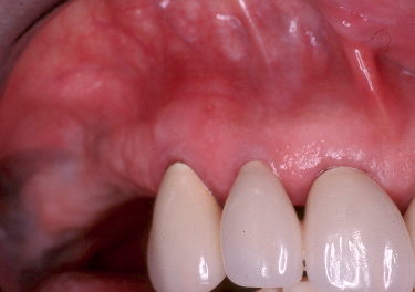
Looking good.
Jeff
Glenn van AsSpectatorWOW Jeff nice tissue result. How is the bone healing , did you take a PA…..I would die to see what is happening there. Is there a reduction in mobility. I am impressed and wouldnt have thought it was possible. That really surprised me and its a cool case. Nice stuff Jeff and man you look young on the new photos (I am in the givin mood as its Xmas time!!)
Glenn
BenchwmerSpectatorHey Glenn,
Good to see you’re in the holiday spirit.
The tooth is solid despite the occlusal tramau. I’ll take a PA and get perio probe readings at the next 3 month recall (that will be 1 year after initial therapy).
I’ve started using digital radiography. I’ll have to figure out how to get the image to the forum.
The PerioLase has created a closed system with the canine. The site was decontaminated, the lining of the fistula tract was removed and a fibrin clot placed in the defect. With a sterile site, no epithelial migration and the building blocks there for regeneration. I’m sure we are getting new attachment (bone, cementum and ligament) to form.
I’m glad you like the new promo pix.
Jeff
JerryDSpectatorJeff,
Ever get any follow-up info on this case?
BenchwmerSpectatorAfter two years, the tooth is still in the mouth. It has never healed completely (maybe due to the tramatic occlusion) It has been re-evaluated at 3 month recalls, a sinus tract has sometimes reappeared, however much smaller and localized than the start. I have repeated the laser therapy and used antibiotic therapy, it clears up. I’ll see where we are at next month.
Jeff
rhuskeySpectatorJeff – Did you do this treatment gratis, or did you charge a fee? If you did, what was your fee, and what insurance code did you file? Thanks Bob Huskey
BenchwmerSpectatorBob,
Some of the time I do it as a favor to the Endodontist and patient.
When billing I use the code D07510 Incision and Drainage Intraoral (My fee 趁)
Jeff -
AuthorPosts