
Forums › Laser Treatment Tips and Techniques › Hard Tissue Procedures › Orthodontic exposure of impacted canine
- This topic is empty.
-
AuthorPosts
-
Glenn van AsSpectatorShoot so tired forgot photos……..
here they are.
Glenn
vinceSpectatorHi Glen,
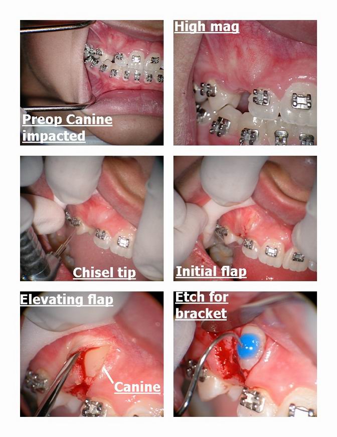
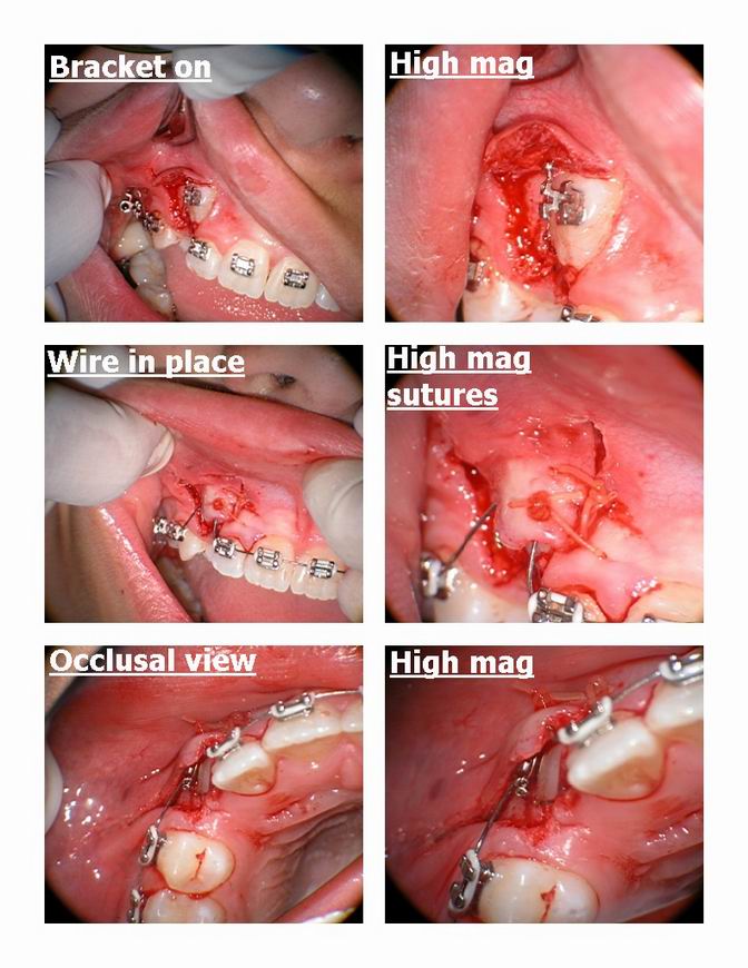
I like to place 2 releasing incisions, one mesial and one distal to the cuspid, and connected at occlusal side with semi-lunar type design.
Make sure you have enough attached gingiva and apically reposition it and suture it down @ cuspid’s CEJ or as close as possible to it. It will come down with the crown and vary rarely requires a graft.Nice case, thanks for sharing.
Vince
Glenn van AsSpectatorHi Vince: thanks for the comments. I infact did place two incisions and a semilunar inicison at the cervical of the teeth.
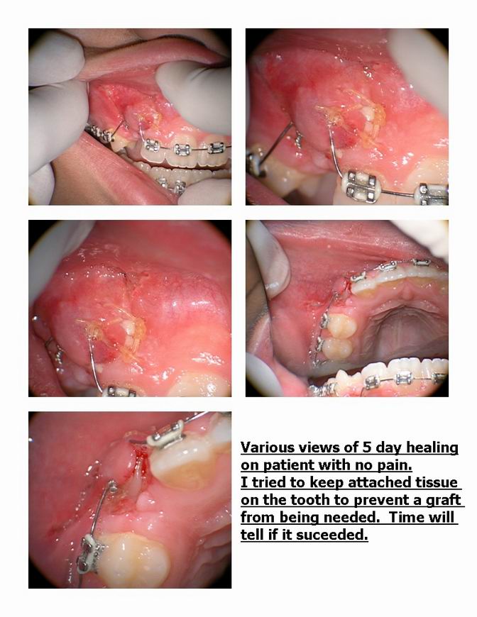
I was leary of the amount of attached tissue and had a heck of a time getting it to place where I did. The tooth is coming quickly so time will tell.
Thanks again………good points. Again surgery , flap design isnt really my strength so each and every time I am learning as I go. I know its pushing the envelope but thats how we all learn.
Glenn
drannetteSpectatorGlenn,
No laser stuff, just a nifty exposure technique:
Recently my daughter had both #6 and 11 exposed. #11 had a dentigerous cyst and was overlying the root of #10 in the depth of the vestibule. #6 was also high up. The oral surgeon laid 2 sizeable flaps from the crest of the ridge, bracketed the teeth with chain attachments, then vertically troughed out the alveolar bone on the buccal to create a path of least resistance for eruption, and fully closed the flaps over the teeth and brackets and sutured at the crest. No windows in the soft tissue.
The sites healed very well and the teeth are moving nicely and should erupt fully into attached gingiva. Cool procedure. I took photos with the surgeon’s camera which should be ready this week. If you are interested I will post them.
Annette Skowronski
Glenn van AsSpectatorHey Annette……..I for one would love to see how he did this, it would be awesome. The flap is great to see how he did it.
Thanks again and take care , hope things are going great.
Cheers
Glenn
drannetteSpectatorGlenn,
Glad you are interested. I’ll post the photos. Just because I felt guilty in not using the laser for the exposures, I removed a fibroma with the DELight today. All the patient and I could say was AWESOME! SHe is returning Saturday for a post op ck. I’ll take healing photos then.Thanks SOO much to everyone who posts (and those that taught at the ConBio seminar) I feel guilty for having so much fun at the office and doing better dentistry to boot!
Annette SKowronski
-
AuthorPosts