
Forums › Laser Treatment Tips and Techniques › Hard Tissue Procedures › YSGG Inlay prep
- This topic is empty.
-
AuthorPosts
-
kellyjblodgettdmdSpectatorToday, I saw a young woman who hates the dental drill. I mean HATES the dental drill. A bur on her tooth to her is like fingernails on the chalkboard.
Anyway, she had an MOD composite placed in #4 roughly 6 months ago. The tooth felt fine prior to the restoration, but has been sensitive ever since the restoration was placed. It was a large composite. Anyway, my exam revealed open gingival margins, probably due to a lack of attention to managing C-factor shrinkage, etc. She got my name off the ALD website, and would prefer that only lasers be used if possible.
Most of the information about how I did it is on the pictures. I’ll try to put up pictures of the pre-op X-ray, if I can figure out how to convert my new digital x-rays to jpeg.
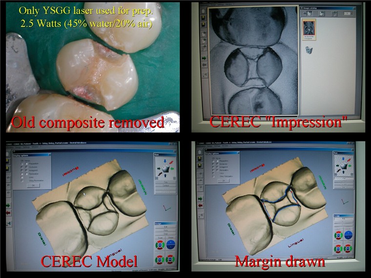
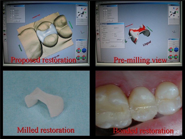
Pt. was very happy. I think the prep could have been a lot smoother, but I was happy with the end result. By the way, the patient was anesthetized, a rubber dam was placed and the entire prep was done under 6.0X mag with the Zeiss Pico microscope.
Kelly
mickey franklSpectatorHi kelly
Great result.
I too combine Cerec and Waterlase preps but usualy get some help from the drill.
Thanks for posting
Mickey
kellyjblodgettdmdSpectatorThanks, Mickey. Do you also find that patients like the lack of vibration?
Glenn on the roadSpectatorGreat documentation Kelly, really very nice result as well.
Anaesthetic (noticed the dam)
Time to do this ( nicely done)
How do you like the Cerec 3d fit with the laser preps which are rougher.CLAP CLAP CLAP……..really well done result.
(Must have been the scope not the clinician right!!)
Just kidding of course……..very good treatment.
Glenn
Lee AllenSpectatorKelly,
Most digital x ray programs allow you to “export” the picture file to email or hard drive where you have a choice of formats like JPEG. See if yours does.The alternative is to open the file in a “photo” program like Adobe has and save it as a JPEG. This program will also allow you to resize for posting here.
Good luck. Nice case. I see that I do not have enough tech toys yet. My lab is very unsettled about the surface texture, but they do it anyway and the results are beautiful. I think in time they will see that it is OK.
kellyjblodgettdmdSpectatorThanks, Glenn. Honestly, I think that the scope aided a lot in making the prep easier. I wasn’t struggling with visibility. Anyway, I did anesthetize the patient. She’s one of those that would prefer to be numb on the way in the front door. No problem with me! The total time from start to finish was just over an hour. So obviously, it helps to be charging for an inlay/onlay if it takes this much time.
And to Lee – I’ll post some photos of surface texture soon. The beauty of this is that you can create whatever you want! Thanks for the help.
Kelly
joegarciaarSpectatorKelly:
Excellent combination!
Laser, Cerec 3D and a very good Dentist.
The new technology in the correct hands makes good works.
whitertthSpectatorKelly,
Great stuff…..The prep is fine as long as the restoration fits, which it looks like it does….What would help u here would be your old enamel hatchets from school or your weidelstadt chisel….U could then the smoothe the preps without rotary if u needed to….Over all…awesome as usual…
kellyjblodgettdmdSpectatorThanks Jose and Ron. Yeah, Ron, I actually do use the enamel hatchets, but honestly, they need a serious sharpening, if you know what I mean. Thanks for the feedback.
In my opinion, the remarkable thing about this case is that I was able to meet all of the patient’s requests by creatively using new technology. She didn’t want vibration and she wanted a restoration that would last longer than the P.O.S. composite she had just had placed – No problem! We can do that!
Kelly
doctorbruSpectatorKelly,
I am looking forward to meeting you and seeing your presentation in Feb. Could you tell me which tip or tips you mostly use for a preparation like this.
Also, where can one learn more about Cerec and the microscope stuff.
Bruce
Glenn van AsSpectatorHey Bruce……..cant answer for the Cerec stuff but a few areas you can find more information about scopes are:
Global Surgical ….. http://www.globalsurgical.com
Academy of Microscope Enhanced Dentistry….http://www.microscopedentistry.com
Microscopes forum..dentaltown…….
http://www.dentaltown.com (look under magnification).Cerec has a board there as well.
Gotta run , hope that helps you a little.
Glenn van As
doctorbruSpectatorThanks Glenn,
What I would really like to know at this moment in time is it possible and practical to have a patient viewing a tv monitor such as the dental chair potato and still have the doctor working thru a microscope. I am having ceiling construction done in my ops and would like to plan on a microscope in the near future.
I’ll ck the sites you listed.
Thanks for posting. Looking forward to meeting you and seeing your presentaion in Feb.Bruce
Glenn van AsSpectatorHi Bruce…….I have a 20 inch Sony Wega in the ceiling a little behind the patient and slightly to the left.
They can then watch TV and the scope.
I know John Kois has both the Couch potato and the scope. Not sure how much he uses the scope….maybe others know.
I like to have the TV slightly off the center (my scope is ceiling mounted as well) so that the patient can still watch while I work. It works ok in my office.
My chairs are slightly off the vertical as well though.
Cya
Glenn
-
AuthorPosts