
Forums › Nd:YAG lasers › General Nd:YAG Forum › Cosmetic Gingivectomy
- This topic is empty.
-
AuthorPosts
-
BenchwmerSpectatorThe following forty something female patient presents with gingival height discrepency on her anterior Maxillary central incisors.
Peroidontal evaluation and probing shows no signs of periodontal disease. Probing shows 3mm to attachment on the facial of #9, another 2mm to sound bone. This should allow me to remove 2mm of gingiva on the facial of #9 without violating biologic width.
Photos before: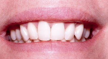

Treatment will consist of Laser Gingivectomy of only tissue covering the clinical crown of #9.
The PerioLase free running, pulsed Nd:YAG, with a contact fiber will be used. LA 4% Citanest infiltrated.
Laser Parameters were 3.0W 20 Hz 150 usec to ablate, bevel and contour the gingival tissues. Duration less than a minute.
Photo immediately after: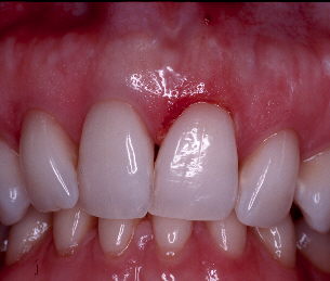
Note precise removal of tissue to expose underlying tooth enamel. The Nd:YAG was chosed for this procedure over the Erb:YAG because of increased hemostatis and not having to protect the root surface during treatment. The laser tip can be directed at a 90 degree angle to the tooth to facilitate tissue removal and final contours without any fear of damage to the underlying tooth or root surfaces.
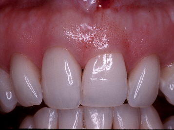
Photo at post-op evaluation at 12 days:
Treatment is complete. Patient is happy.
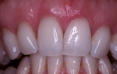
Before/After:
(Edited by Benchwmer at 11:36 am on May 22, 2004)
(Edited by Benchwmer at 11:39 am on May 22, 2004
(Edited by Benchwmer at 11:46 am on May 22, 2004)
ASISpectatorHi Jeff,
Well handled and documented case. A simple procedure can make a world of difference on an asymmetrical gingival contour even though her smile line barely displays it.
Cheers.
Andrew
Glenn van AsSpectatorHI Jeff: I like this one alot for alot of reasons, the fact that you got great photos. The fact that the case was beautifully handled and subtle things like the lack of char are all wonderful.
In addition, I think it is neat to see how there was slight rebound in this case.
I wonder if we did erbium osseous (open or in this case a mm of closed flap) if we could get the rebound to not occur.
I would estimate that 75% of the original amount of removal stayed in place.
I think from your case and ones I have done as to whether I will tell patients its a 2 phase treatment with soft tissue first and if needed hard tissue after wards.
Perhaps it can be combined if we are adequate in our probing depths…….
Neat case Jeff, and a big big service to your patient.
CLAP CLAP CLAP……..love to see various lasers for different treatments I learn each time I see em.
Cya
Glenn
BenchwmerSpectatorGlenn,
25% rebound? May be camera angles.
Notice how I didn’t touch the mid-line papilla. I you laid a flap you would be able to see the height of bone, use the Erbium to crown lengthen, but you are adding the variable of healing after a flap, added expense, post-op complications.
In this case the worse case scenario is the tissue doesn’t heal to the height after laze. If, the patient doesn’t like the results, it is a 10 minute procedure to remove more tissue if probing and bone sounding merit the attempt or if not, then flap/remove bone.
Case selection determines the “How to”.
I’ll post a soft tissue Erbium use later.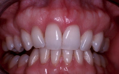
Jeff
AnonymousGuestCan anyone tell me why this extra tissue is there to begin with? Jeff, did you probe the sulcus after removing the tissue? How did you decide you could remove 2mm, i.e. how did you determine the 3mm BW after Tx would be enough?
Also, if you did flap and didn’t touch the bone interproximally, couldn’t you expect the papilla to return to normal position?
Sorry for all the questions, just trying to understand this BW and anterior cosmetic procedures better.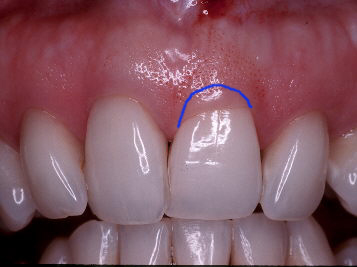
BenchwmerSpectatorRon,
These are my thoughts on biologic width. “Studies” (I don’t footnote quickly like you, I learned fron my DentalTown posting, you can reference the responses to my laser cases on DT case studies) show you need 2.6mm of biologic width. I measured 3mm from crest of the gingiva to the base of the pocket, under LA I sounded bone at 5mm from the gingival crest. That gives me 5.0mm minus 2.6mm or 2.4mm of free gingiva I can excise without violating BW. I measured 2mm and used a perioprobe to create bleeding points in 5 or 6 right (where you placed the line). This lets me connect the dots and remove a finite measured amount of tissue.
I’ll try to get you a couple of good articles to read.
I won’t probe the area for 6 months. Will expext less than 2mm of probe depth.
Jeff
Andrew SatlinSpectatorRon,
To answer part of your question, In my experience (12 years of perio sx) if you completely reflect the papilla you will lose soft tissue height even if you do not remove interproximal bone.
There are alot of factors involved particularly tissue thickness but when osseous recontouring is required on cases like this, I split the papillae and leave as much interprox tissue as possible.
This gives you good acces to the buccal bone and you can delicately ramp it interproximally if you need to.
Vertical matress suture to minimize pressure on the papillary height.
Try it sometime (or refer your patient to me!!!)–just joking
See you all
Andy
Dan MelkerSpectatorAnswer to Ron’s question,
Cause of tissue position-
1. traumatic injury when this patient was young(#9 intruded)
2. tooth in lingual version
These 2 factors may effect the outcome of this beautifully done procedure.
I think why Glenn thought there was rebound of tissue was because #9 is intruded. Just a thought.
Thanks,
Danny(Edited by Dan Melker at 9:16 am on May 23, 2004)
whitertthSpectatorJeff,
Very nicely done…..
Questions….I want to make sure I understand u …You probed the sulcus and got a measurement, then went thru the attachment under local got another measurment(Bone_) and subtracted first from second to know how much tissue u could remove…Is that correct?
Now a quick comment … Mind u a great result…But I beleive that u could have achieved as nice a result with the Waterlase with minimal bleeding and done it without local especially with my DRKLIQUID topical…. Just a thought but still outstanding case…
Glenn van AsSpectatorThat brings me to a question I have………
Ron I want to know what topical I can get here in Canada that I can order and it will get through the border.
I have a frenum to cut soon on a child and mom wants no local.
ANy ideas Norman, Andrew or my Canadian compadres.
Glenn
ASISpectatorHi Glenn,
I have used EMLA mostly along with Hurricaine topical spray and adequate water irrigation with the 980nm diode. With all of the above going on, and if greater comfort is still required to complete the treatment, I will have no hesitation to place judicious drops of local as needed.
As you know, some parents are the greatest cause of discomfort in terms of anxiety that they induce, as much if not more so than the procedure itself….
Cheers,
Andrew
BenchwmerSpectatorThe same results could be achieved using different tools.
If I’m going to soung to bone with a perio probe, patients prefer LA, a few drops of 4% Citanest infiltrated into the tissues is all that is needed. I place a topical before injecting. I have purchased the DrKLiquid.
I just find very limited uses for it as a replacement for LA.
The biggest advantage of using the Nd:YAG in this type of case is you don’t have to protect the root surface from the laser tip, the procedure can be done with the fiber directly pointed at the tissue, the tissue can be ablated, the margins beveled, tissue tags removed without harming the root surface, with an Erbium I would have to use the tip parrallel to the tooth. Plus better hemostatis w/ Nd:YAG.
I have the choice of lasers.
Why is it such a big deal for Waterlase users not to use LA?
I was at a Preview session last month, and saw the Waterlase user, firing the fiber into the sulcus surrounding a Mandibular molar, 4 different points, took about 3 minutes, all for a buccal pit that would not have been painful if the Erbium would have been used on the tooth directly.
What effect does the laser energy have on attachment?
Jeff
Glenn van AsSpectatorHi Andrew……..where can I get EMLA topical.
Glenn
ASISpectatorHi Glenn,
Any pharmacy. No Rx required.
Andrew
whitertthSpectatorGlen,
contact me offlist and lets seeif we can come up with [email=”something…whitertth@aol.com”]something…whitertth@aol.com[/email]
516 521 0394 cell -
AuthorPosts