
Forums › Laser Treatment Tips and Techniques › Hard Tissue Procedures › Diagnosis of Occlusal caries
- This topic is empty.
-
AuthorPosts
-
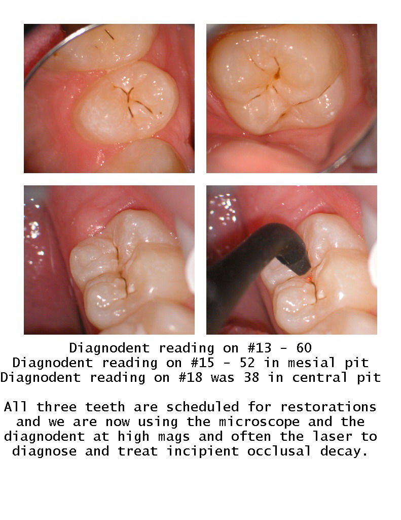
Glenn van AsSpectatorHere is what we are doing now. We use the microscope at 10X power with air from the triplex syringe to dry of the tooth and evaluate areas that we think may be decay. We see halos, stain and cavitated and decalcified areas.
The diagnodent gives us a reading , and usually anything over 25 is decay into dentin.
HEre are a couple of non cavitated lesions I saw today in one patient. These are the types of cavities I see now with Fluoride in the toothpastes making caries so hard to daignose.
Glenn
ASISpectatorHi Glenn,
Nice diagnostic sequence. A powerful combination of tools to confirm needed treatment.
I guess I ‘ll have to breakdown to get that Diagnodent to complete my diagnostic armamentarium, although I have treated many of these and found active carious lesions without it.
You continue to motivate me to strive for perfection, Glenn. Thanks again.
Andrew
whitertthSpectatorAndrew,
The Diagnodent is amust and goes hand in hand with laser dentistry….Dont walk , run get a diagnodent. It will pay for itself in 1 month….
dkimmelSpectatorGlenn man your asking for trouble. I got slammed by a new patient today. I Dx about 10 cavities on this 40 Y/O female. She was changing dentist because it was just too far to go and see him. Long story short , she goes back to see him. He sends her back to me with his X-ray , to tell me he sees no decay. I look at his film and sure enough no decay just an occulsal alloy. She wants to know whats up. I have to pull out her photos and show her the open margin on the mesial , the color change and the crack. So she is back to trusting me again. Now she tells me her tooth hurts. We open it up and the mesial has a good size cavity and the lingual cusps and the mesial buccal cusp is fractured. The fracture from the lingual cusp goes torward the distal and through the pupal floor. Endo time.
This lady has been seen every 6 mo for the last 10 years. It is highly probable that this could have been picked up before it came to endo. The Diagnodent and magnifaction are far superior to X-rays and the explorer for detecting caries.
David
marc andre gagnonSpectatorhi
I am a diagnodent user since more than 3 years
All the hygienists in the office have a diagnodent in their operatories.We use it on each exam since the beginning.They mark the decay point on the dental chart and I do the filling exactly where they mark it.
Be sure that a diagnodent will pay for itself in 1 month….
Maybe like david said that you can have problem with new patient but I’m sure of the quality of the job we do each time we use diagnodent.
2thlaserSpectatorGlenn,
This is stuff for a lecture at the World Congress of Microdentistry. I wish you would consider being there with us this next August in San Fransisco, and present magnification, AND the diagnodent. Nice stuff. I see the same things with my loupes, (just not as big!) 6x vs. 10x! Quite a difference I might say!
Mark
Glenn van AsSpectatorHi Mark: if I get an official invitation and confirmation that I will be invited to speak I will come. I am so busy these days lecturing that it is pretty well impossible for me to attend meetings for only CE purposes.
Having said this if you so invite me to speak I will then combine my lecture with staying for the whole meeting and seeing what the WCM has to offer.
I value the experience that you, Graeme, Stewart and a few others show consistently, and think that I could do a fun lecture on something.
Let the powers be know that I would be interested in presenting on any of the following:
Documentation of microdental procedures through the D.O.M.
Magnification and microdental preparations.
The dynamic duo in dental decay diagnosis: The D.O.M. and the Diagnodent…….da da!!
(I love alliteration )
Cya
Glenn
2thlaserSpectatorGlenn,
I know I am speaking for the others when I say we would be honored to have you speak. I will contact Kim, Stu, and Joe Whitehouse our president. I am sure, absolutely sure they will have you speak. There are no honorariums…which is nice. We speak and teach because of our desire for the advancement of microdentistry. I will be in touch with you personally soon.
From Chicago!
MarkPS…you going to ADA? next week?
Glenn van AsSpectatorYes Mark , I have two lectures on scopes…….one on the 23rd and one on the 26th.
Hope to see you around.
Drop by either the Global booth or the Hoya booth on the 24th or 25th as I will be helping them out then.
Gotta go get my lectures done.
Cya
Glenn
Samuel MossSpectatorAndrew,
I just want to add that my staff was a little wary when we first started using the Diagnodent a little over a year and a half ago. One patient left, went to another dentist who told her that I probably had to pay for all my machines but he was sure there was no decay. I had known her for almost 20 years. So, I used it on my staff and lo, my hygenist had some readings in the 50s. I decided to photograph my way through her fillings. On her lower 2nd molar, I had to end up using anesthesia due to the fact that the lesion was so large. Of course, you could not detect it with x-ray or the braille method ( explorer). Long story short, when she uses the Diagnodent on patients, she tells her story and can show the photos of how nothing became a big “something”.
Just thought I would share
Mossman
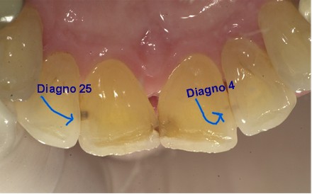
dkimmelSpectatorTo trust the Diagnodent or not? Today I had a tough time trusting the Diagnodent. Two lesions one read 25 and the other 4. The 25 is a no brainer but the 4 ? I can see the lesion and it says “lase me”. What do you think?
(Edited by dkimmel at 5:55 pm on Oct. 21, 2003)
SwpmnSpectatorDave:
What is your history with this patient? Is this a new patient or are these lesions you’ve observed for awhile?
I don’t presently use the Diagnodent. However, at twelve years out have observed many lesions at your “4” setting that NEVER change. The other three definitely need to be restored.
My proposal is that the “4” lesions are arrested and your Diagnodent is right on target.
Just an opinion from someone who has never clinically used the Diagnodent,
Al
dkimmelSpectatorAl, She is a new patient. The photo’s are from her NP exam. I think you are right but I am itching to use that laser some more!

DAvid
drcamSpectatordavid,
i’ve been a diagnodent user for about 3 years and generally only use it for occlusal caries detection. My understanding is that interproximal lesions are hard to assess with it depending on how much enamel is overlying the caries. I would probably observe those lesions and try to remineralise them. The other 3 are obviously into dentine however and are waiting for some ablation. From a patient education perspective i use graeme milicich’s cd rom to get an instant 2nd opinion which boosts your credibility.Cheers cam
-
AuthorPosts