
Forums › Laser Resources › Laser Endo Related Literature › Negative article on Er,Cr:YSGG and endo disinfection
- This topic is empty.
-
AuthorPosts
-
Glenn van AsSpectatorHere is the article, what say ye people.
J Am Dent Assoc, Vol 137, No 1, 67-70.
© 2006 The American Dental Association
This ArticleAbstract
Full Text (PDF)
Alert me when this article is cited
Alert me if a correction is postedServices
Similar articles in this journal
Alert me to new issues of the journal
Download to citation managerGoogle Scholar
Articles by Jha, D.
Articles by Hasselgren, G.PubMed
Articles by Jha, D.
Articles by Hasselgren, G.
RESEARCHInability of laser and rotary instrumentation to eliminate root canal infection
D. Jha, DDS, A. Guerrero, DDS, T. Ngo, DDS, A. Helfer, DDS, MSD and G. Hasselgren, DDS, PhD
ABSTRACT
TOP
ABSTRACT
MATERIALS AND METHODS
RESULTS
DISCUSSION
CONCLUSION
REFERENCESBackground. The authors evaluated the antibacterial effectiveness of laser instrumentation and rotary instrumentation of anterior, single-rooted teeth infected with Enterococcus faecalis.
Methods. The authors divided 35 infected samples into five groups: Group A: inoculation, laser, 17 percent ethylene-diamine-tetra-acetate (EDTA), 2.5 percent sodium hypochlorite (NaOCl) (n = 10); Group B: inoculation, laser, 17 percent EDTA, sterile saline (n = 10); Group C: inoculation, rotary, 17 percent EDTA, 2.5 percent NaOCl (n = 10); Group D: inoculation, no instrumentation (positive control) (n = 5); Group E: no inoculation, no instrumentation (negative control) (n = 5). They sampled and incubated dentin shavings from each canal for bacterial growth.
Results. In Group A, eight tubes were positive for bacterial growth. In Group B, 10 tubes were positive for bacterial growth. In Group C, six tubes were positive for bacterial growth. In Group D, all of the tubes were positive for bacterial growth. In Group E, no tubes showed bacterial growth. The Fisher exact test showed no significant differences among groups A, B and C.
Conclusion. Neither the laser nor the rotary instrumentation was able to eliminate endodontic infection.
Clinical Implications. Although lasers have been presented as high-tech tools for disinfecting root canals, the laser was ineffective in this study.
Key Words: Laser; root canal disinfection; root canal instrumentation
Various types of lasers have been studied in an attempt to develop improved treatment methods in health care. In 2002, the U.S. Food and Drug Administration (FDA) approved the erbium, chromium: yttrium, scandium, gallium, garnet (Er,Cr:YSGG) laser for use in conventional and endodontic therapy. The Er,Cr:YSGG crystal generates photons through a fiber-optic cable delivery system terminating in a handpiece with a sapphire crystal that is bathed in air-water spray.1 The Er,Cr:YSGG laser emits an invisible beam in the infrared range of 2.79 micrometers, coupled with a nonabsorbing light source that serves as a pointer for the working laser.2
The photon energy for this system interacts with the water spray; the maximum energy is delivered 1 to 2 millimeters from the sapphire tip. The laser ablates tissue in less than a 5-mm range.1
A review of the literature revealed that investigations of the use of the Er,Cr:YSGG laser in endodontics exist, but they are limited in number and scope. Some of these studies have addressed the response of pulpal and canal walls to irradiation temperature, the morphology of laser-instrumented root canal walls, and the presence or absence of the smear layer after laser treatment.3–5 Ishizaki and colleagues’6 study found that the Er,Cr:YSGG laser caused a rise of 8 C on the root surface and that it was efficient in removing the smear layer without carbonizing or melting the dentin.
Infection is the cause of pulpal and periradicular diseases.7 Bacteria and their byproducts are considered to be the primary etiologic agents of pulpal necrosis and periapical lesions. Therefore, the elimination of bacteria and their byproducts is one of the most important steps in endodontic treatment.8,9 A few studies have dealt with using lasers to disinfect root canals, but their methods and materials were not sufficient. For example, Schoop and colleagues10 concluded that all of the lasers that they investigated (Er,Cr:YSGG, neodymium-doped yttrium aluminum garnet and erbium substituted:yttrium aluminium garnet) are suitable for disinfection of even deep layers of dentin. However, the authors only allowed bacteria to infect the dentin for four hours before using the lasers.
Microbiological studies have shown that in failed endodontic therapy, the microbial flora often consists of a single species of predominantly gram-positive organisms. The most commonly recovered isolates were from the bacteria Enterococcus faecalis.11,12
In reference to microbiology, no studies have examined the ability of Er,Cr:YSGG laser to disinfect root canals during endodontic treatment. Therefore, we conducted this study to compare the ability of an Er,Cr:YSGG laser (WaterLase, Biolase Technology, San Clemente, Calif.) to disinfect the root canals of extracted human teeth infected with E. faecalis with the ability of the nickel titanium Profile 0.06 taper rotary file series (Dentsply Tulsa Dental Products, Tulsa, Okla.).
MATERIALS AND METHODS
TOP
ABSTRACT
MATERIALS AND METHODS
RESULTS
DISCUSSION
CONCLUSION
REFERENCESWe used 40 extracted, permanent, anterior, single-rooted teeth in our study. We decoronized and instrumented the teeth with International Organization for Standardization (ISO) nos. 10, 15 and 20 with hand files to the teeth’s working length. We determined working lengths by inserting a no. 10 hand file into the root canal until it was visible at the apical foramen and then subtracted 1 mm from that length. We stored the teeth in sterile saline (0.9 percent sodium chloride) for three days and then autoclaved for them 20 minutes at 121 C.
Infection and instrumentation. We used facultative anaerobic bacteria (E. faecalis ATCC no. 27792 [American Type Culture Collection, Manassas, Va.]) in our experiment. We rehydrated a freeze-dried culture pellet with 0.5 milliliters of phosphate-buffered saline solution, streaked onto Brucella blood agar (Anaerobe Systems, Morgan Hill, Calif.) plates and incubated them at 37 C in anaerobic incubator boxes.
We saw colony growth after three days. We used an inoculating loop to take single colonies from the plates and used them to inoculate 7 mL of thioglycollate (THIO) medium broth (Anaerobe Systems) in sterile test tubes. We then randomly divided the autoclaved teeth into three experimental groups of 10 teeth each (Groups A–C), one positive control group of five teeth (Group D) and one negative control group of five teeth (Group E). We infected the dentin according to the method described by Haapasalo and Orstavik.13 We placed the teeth to be infected (those from Groups A–D) in separate test tubes containing THIO broth inoculated with E. faecalis. We then placed the test tubes in anaerobic incubator boxes and incubated them at 37 C for 21 days. Every third day, we replaced 3 mL of the inoculated THIO broth with 3 mL of fresh THIO broth. At the end of the incubation period, all the test tubes showed positive turbidity for bacterial growth. We removed the infected teeth from their test tubes, rinsed them with sterile saline and treated them as follows.
We laser instrumented the 10 infected teeth in Group A following the steps outlined in the manufacturer’s instructions. The firing range settings for the Z2, Z3 and Z4 laser tips were 1.50 watts, 34 percent air and 24 percent water. We irrigated the canal with 2.5 percent sodium hypochlorite (NaOCl) after instrumentation with the Z2 and Z3 laser tips. After instrumentation with Z4 laser tip, we irrigated the canal with 1 mL of 17 percent ethylene-diamine-tetra-acetate (EDTA) for one minute, followed by a rinse of 2.5 percent NaOCl. The total volume of 2.5 percent NaOCl we used to irrigate each tooth was 3 mL via a plastic syringe with a 23-gauge needle.
We laser instrumented the infected teeth in Group B in almost the same manner as we instrumented the infected teeth in Group A. The only difference was that we used sterile saline instead of 2.5 percent NaOCl to irrigate the canal. The firing range of settings for the Z2, Z3 and Z4 laser tips were 1.50 W, 34 percent air and 24 percent water. We irrigated the canal with sterile saline after instrumentation with the Z2 and Z3 laser tips. After instrumentation with the Z4 laser tip, we irrigated the canal with 1 mL of 17 percent EDTA for one minute, followed by a rinse with sterile saline. The total volume of sterile saline we used to irrigate each tooth was 3 mL via a plastic syringe with a 23-gauge needle.
We instrumented the infected teeth in Group C with the taper rotary file series in sizes ISO nos. 20, 25, 30, 35 and 40. We irrigated the canal with 2.5 percent NaOCl after instrumentation with each rotary file. After instrumentation with the no. 40 rotary file, we irrigated the canal with 1 mL of 17 percent EDTA for one minute, followed by a rinse with 2.5 percent NaOCl. The total volume of 2.5 percent NaOCl we used to irrigate each tooth was 3 mL via a plastic syringe with a 23-gauge needle.
After 21 days of incubation at 37 C, the infected teeth in the positive control group (Group D) were not instrumented. Instead, we collected dentin shavings from the teeth and incubated them for 21 days at 37 C in sterile test tubes containing 7 mL of THIO broth.
We did not inoculate teeth in the negative control group (Group E), but we did incubate them for 21 days at 37 C in sterile test tubes containing 7 mL of THIO broth.
We analyzed the differences between groups using the Fisher exact test.
Dentin shaving collection. Immediately after laser or rotary instrumentation of teeth from Groups A to C, we rinsed the teeth from all five groups (A–E) with sterile saline. We collected dentin shavings, using the methods described by Gomes and colleagues.14 For each tooth, we obtained dentin shavings using sequential sterile Gates-Glidden (GG) burs nos. 2, 3 and 4 at low speeds. We used GG bur no. 2 until it went to working length and burs nos. 3 and 4 until we felt resistance. After instrumentation with the GG burs, we used an ISO no. 25 hand file until it went to the working length of each canal. We then suspended the dentin shavings with sterile saline within the canal. We introduced sterile paper points into the canals to collect the dentin shavings and transferred the shavings into separate test tubes containing fresh THIO broth. We placed the test tubes in anaerobic incubator boxes and incubated them at 37 C. After 72 hours, we examined the test tubes for microbial growth and checked for medium turbidity. To confirm the presence of E. faecalis, we plated all of the broths that resulted in turbidity on Brucella blood agar plates.
RESULTS
TOP
ABSTRACT
MATERIALS AND METHODS
RESULTS
DISCUSSION
CONCLUSION
REFERENCESOf the 10 test tubes from Group A, two showed no turbidity, while eight were positive for bacterial growth (Figure). All 10 of the test tubes from Group B were positive for bacterial growth. In Group C, four test tubes showed no turbidity, while six test tubes were positive for bacterial growth. All five of the test tubes from Group D were positive for bacterial growth. None of the five test tubes from Group E had bacterial growth. When we conducted statistical analyses using the Fisher exact test, we found that the differences between Groups A, B and C were not statistically significant.
View larger version (50K):
[in this window]
[in a new window]
Figure. Distribution of samples positive and negative for bacterial growth. NaOCl: 2.5 percent sodium hypochlorite.After we inoculated Brucella blood agar plates with broth from the turbid test tubes, we found that the resulting colonies had similar growth patterns to those of E. faecalis stock plates.
DISCUSSION
TOP
ABSTRACT
MATERIALS AND METHODS
RESULTS
DISCUSSION
CONCLUSION
REFERENCESIn 2002, the FDA approved the Er,Cr:YSGG laser for use in conventional and endodontic therapy. FDA approval of the Er,Cr:YSGG laser for use in endodontic therapy does not mean that the laser is effective in disinfecting root canals. Because no studies have examined Er,Cr:YSGG laser instrumentation in reference to microbiology, we were interested in comparing the antimicrobial effects of laser instrumentation to that of rotary instrumentation. We used rotary instrumentation with 25 percent NaOCl irrigation as the comparison technique because it is common in conventional endodontic treatment.
In our study, we chose E. faecalis because it commonly is recovered from teeth in endodontic cases that failed.11,12 We incubated the infected teeth for 21 days to ensure adequate bacterial penetration into dentinal tubules. Haapasalo and Orstavik13 showed heavy infection of E. faecalis to a depth of 400 µm in tubules after 21 days.
Studies have shown that mechanical instrumentation alone and using sterile saline to irrigate the canals significantly reduces the number of bacteria in infected root canals. According to studies by Byström and Sundqvist,15,16 however, a completely negative culture cannot be obtained without the use of antibacterial irrigation agents. The results of Group B, in which 10 of 10 test tubes were positive for bacterial growth, corroborate the findings of Byström and Sundqvist.15,16 Likewise, the bacteria-free test tubes from Groups A and C support the use of antibacterial irrigation as a key component in achieving a bacteria-free root canal.
Lasers have been presented as a revolutionary technology and as the future of endodontics.17 The results of this study do not support this concept. It appears that no instrumentation technique alone can rid a canal of infection.
CONCLUSION
TOP
ABSTRACT
MATERIALS AND METHODS
RESULTS
DISCUSSION
CONCLUSION
REFERENCESNeither rotary nor Er,Cr:YSGG laser instrumentation was able to eliminate an E. faecalis infection in root canals when 25 percent NaOCl was used as an irrigation solution. Further, the laser was completely ineffective in disinfecting root canals when sterile saline was used as an irrigation solution.
FOOTNOTES
Dr. Jha is a clinical instructor, Division of Endodontics, School of Dental and Oral Surgery, Columbia University, 630 West 168th St., PO Box, 20 (PH7E-117), New York, N.Y. 10032, e-mail “dm762@Columbia.edu “. Address reprint requests to Dr. Jha.
Dr. Guerrero was a postdoctoral student, Division of Endodontics, School of Dental and Oral Surgery, Columbia University, New York City, when this article was written. He now maintains a private practice in Santa Monica, Calif.
Dr. Ngo was a postdoctoral student, Division of Endodontics, School of Dental and Oral Surgery, Columbia University, New York City. He now maintains a private practice in San Francisco.
Dr. Helfer is a clinical associate professor, Division of Endodontics, School of Dental and Oral Surgery, Columbia University, New York City.
Dr. Hasselgren is the director, Division of Endodontics, School of Dental and Oral Surgery, Columbia University, New York City.
REFERENCES
TOP
ABSTRACT
MATERIALS AND METHODS
RESULTS
DISCUSSION
CONCLUSION
REFERENCESRizoiu IM, Eversole LR, Kimmel AI. Effects of erbium, chromium: yttrium, scandium, gallium, garnet laser on mucocutanous soft tissues. Oral Surg Oral Med Oral Pathol Oral Radiol Endod 1996;82:386–95.[Medline]
Goodis HE, Pashely D, Stabholtz A. Pulpal effects of thermal and mechanical irritants. In: Seltzer and Bender’s dental pulp. Hargreeves KM, Goodis HE, eds. Carol Stream, Ill.: Quintessence; 2002:371–88.
Matsumoto K. Lasers in endodontics. Dent Clin North Am 2000;44(4);889–906.[Medline]
Goya C, Yamazaki R, Tomita Y, Kimura Y, Matsumoto K. Effects of pulsed Nd:YAG laser irradiation on smear layer at the apical stop and apical leakage after obturation. Int Endod J 2000;33:266–71.[Medline]
Eversole LR. Rizoiu IM. Preliminary investigations on the utility of an erbium, chromium YSGG laser. J Calif Dent Assoc 1995;23(12):41–7.[Medline]
Ishizaki NT, Matsumoto K, Kimura Y, Wang X, Kinoshita J, Okano SM. Thermographical and morphological studies of Er,Cr:YSGG laser irrradition on root canal walls. Photomed Laser Surg 2004:22(4)291–7.[Medline]
Kakehashi S, Stanley HR, Fitzgerald RJ. The effects of surgical exposures of dental pulps in germ-free and conventional laboratory rats. Oral Surg Oral Med Oral Pathol 1965;20:340–9.[Medline]
Bergenholtz G. Micro-organisms from necrotic pulp of traumatized teeth. Odontol Revy 1974;25:347–58.[Medline]
Sundqvist G. Bacteriological studies of necrotic dental pulps (dissertation). Umeå, Sweden: Umeå University; 1976.
Schoop U, Kluger W, Moritz A, Nedjelik N, Georgopoulos A, Sperr W. Bactericidal effect of different laser systems in the deep layers of dentin. Lasers Surg Med 2004;35:111–6.[Medline]
Molander A, Reit C, Dahlen G, Kvist T. Microbiological status of root-filled teeth with apical periodontitis. Int Endod J 1998;31:1–7.[Medline]
Sundqvist G, Figdor D, Persson S, Sjögren U. Microbiologic analysis of teeth with failed endodontic treatment and the outcome of conservative re-treatment. Oral Surg Oral Med Oral Pathol Oral Radiol Endod 1998;85(1):86–93.[Medline]
Haapasalo M, Orstavik D. In vitro infection and disinfection of dentinal tubules. J Dent Res 1987;66(8):1375–9.[Abstract/Free Full Text]
Gomes BP, Souza SF, Ferraz CC, et al. Effectiveness of 2 percent chlorhexidine gel and calcium hydroxide against Enterococcus faecalis in bovine root dentin in vitro. Int Endod J 2003;36:267–75.[Medline]
Byström A, Sundqvist G. The antibacterial action of sodium hypochlorite and EDTA in 60 cases of endodontic therapy. Int Endod J 1985;18:35–40.[Medline]
Byström A, Sundqvist G. Bacterial evaluation of the efficacy of mechanical root canal instrumentation in endodontic therapy. Scand J Dent Res 1981;89:321–8.[Medline]
Chen WH. YSGG laser root canal therapy. Dent Today 2002;21(5):74–7.[Medline]
This Article
Abstract
Full Text (PDF)
Alert me when this article is cited
Alert me if a correction is postedServices
Similar articles in this journal
Alert me to new issues of the journal
Download to citation managerGoogle Scholar
Articles by Jha, D.
Articles by Hasselgren, G.PubMed
Articles by Jha, D.
Articles by Hasselgren, G.
HOME HELP FEEDBACK SUBSCRIPTIONS ARCHIVE SEARCH TABLE OF CONTENTS
dkimmelSpectatorJust from a quick read I have 3 concerns about this article
The first of which is the laser used. What controls do they have to indicate the the energy level they are using are correct. I have learned form the periolase and its built in meter , that what you think you have is not always what you get. So what control do they have to show that this laser is working correctly.
The second is thier confirmation that the bacteria they found present is the E. faecalis they are using is done by seeing if the colonies appear the same on the Brucella blood agar.It is a very simple test to determine if what was present was indeed E. faecalis. The agar they are using can support the growth of other bacteria that may appear simplier to E. faecalis. Heck even a Gram stain should have been done. If you consider the difficulty in doing this study and maintaining a sterilty control , confirmation should have been part of the controls. A Negative control alone is not enough.
Third is a question of bioburden.
We placed the teeth to be infected (those from Groups A–D) in separate test tubes containing THIO broth inoculated with E. faecalis. We then placed the test tubes in anaerobic incubator boxes and incubated them at 37 C for 21 days. Every third day, we replaced 3 mL of the inoculated THIO broth with 3 mL of fresh THIO broth. At the end of the incubation period, all the test tubes showed positive turbidity for bacterial growth. We removed the infected teeth from their test tubes, rinsed them with sterile saline and treated them as follows.Excuse me but 21 days in THIO broth refreshed every 3days. First off when you look at antimicrobial activity you ALWAYS quantify the number of bacteria being used in the study. In 24 hrs hours the numbers of bacteria in THIO are hugh. In 21 Days there would be so many that in NO way would this even come close to the bioburden seen in a clinical setting. It would also mean that the tech doing this would need to be extremely careful not to cross contaminate samples , instruments etc…. More then that the bioburden within the canals/tublules would be so high that there would be no way to get 100% kill. You would have to get 100% kill the way they did the test in order to get a neg result. Again they incubated the samples for 72 hrs before plating. Just one bacteria present would give a postive test.
Frankly I would find it hard to believe this study could get published in a Journal other then a dental one. It is poorly designed. If the had done colony counts to determine the bioburden at the start of the study and if they had quantified the bacteria # in the samples at the end of the study, it would have been a much better study.
AnonymousGuestTwo other potential problems-
1.They laser instrumented according to manufacturer directions (I assume ala Chen by the reference?)
: Photomed Laser Surg. 2005 Apr;23(2):196-201. Related Articles, Links
Efficacy of root canal preparation by Er,Cr:YSGG laser irradiation with crown-down technique in vitro.
Ali MN, Hossain M, Nakamura Y, Matsuoka E, Kinoshita J, Matsumoto K.
Department of Endodontics, Showa University School of Dentistry, Tokyo, Japan. thebestdentist@yahoo.com
OBJECTIVE: The purpose of this study was to compare the efficacy of Er,Cr:YSGG laser in root canal preparation and its effectiveness in removing debris and smear layer with the efficacy of the conventional hand instrument, in vitro. BACKGROUND DATA: Recently, the use of the Er,Cr:YSGG laser device in root canal preparation has been excepted in the dental clinic. However, there have been no published reports on root canal preparation by using the Er,Cr:YSGG laser irradiation. MATERIALS AND METHODS: A total of 40 straight root canals were prepared by an Er,Cr:YSGG laser at a output power of 2 W, and 40 canals were shaped by using the K file under irrigation with NaOCl and H2O2 by using the crown-down technique (control). The achievement degree of root canal preparation and debris score was morphologically investigated. RESULTS: The results showed that great improvement in the cleanliness of the walls was found using the laser technique as compared with the control technique; a significant decrease in smear layer or debris was also recognized in laser-prepared canals (p < 0.01). But canal preparations with the laser device sometime result in ledge, zipped, perforation, or over-instrumentation. CONCLUSION: The results demonstrated that root canal preparations with the laser device were significantly worse than in the control group. Further development in laser device and technique are required to ensure its success in root canal preparation.
PMID: 15910186 [PubMed – indexed for MEDLINE]
2. could part of the problem be the laser energy was suffiently absorbed in the water spray component to prevent adequate disinfection? Another good reason to choose a wavelength that water is invisible to, eg nd:YAG
2thlaserSpectatorTime to chime in here a bit. For those of you who don’t know, I have been doing research for over a year now on endo with the YSGG wavelength with Dr. DiVito in Scottsdale, AZ. We chose to do our research with our own money, and scientists so as not to have our results skewed in favor of a manufacturer of any kind, or a school’s research agenda. David knows this. First, David is exactly right in his description of the research flaws. There is a ton more to extrapolate on here, but with non-disclosures in place, you guys have to trust me when I say the research has a “taint” in it. Secondly, I assume that the people who performed the research were NOT trained on the laser to my knowlege, and therefore probably do NOT know the protocols in using the laser correctly in a root canal system.
Dr. DiVito and I have demonstrated the proper techniques, which David has seen, in how to use the laser in a canal, but have furthered the research even more so with science that I can only show you a part of at this time.
I will make some generalizations here, that seem logical, but criticism is welcome…
If the smear layer can be removed, and no bacteria is present under Scanning Electron Microscopic view, can it be assumed that the canal(s) have been effectively decontaminated? Now I didn’t say sterilized here.Example One:
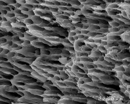
Clean, “at attention” dentin in the canal. No smear layer present, prepared with Er,Cr:YSGG laser, 1.5w, 34%air, 24% water, canal prepared with a MZ-2 fiber at 20hz, laser energy is activated on the outstroke only, 4 outstrokes of laser energy, about 8 seconds exposure time on each outstroke, with no EDTA, or NaOCl just laser energy and the water from the laser. Bacteria present in SEM?
Number 2:
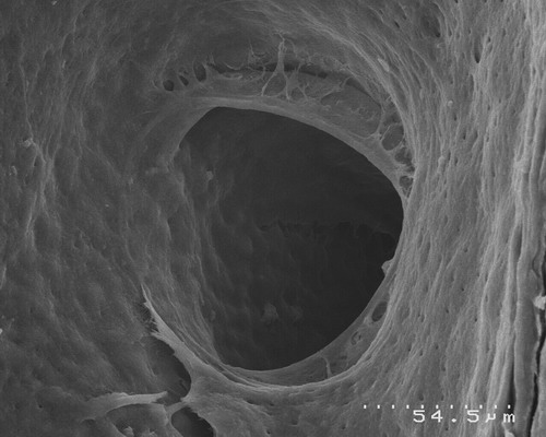
Clean lateral canal, no smear layer, same laser parameters as above. Can a file with NaOCl, EDTA, 30 MINUTES of irrigation time do this? Yes, but I believe not as effectively as Dr. DiVito and I have begun to demonstrate. Can a file negotiate a lateral canal? I haven’t seen it yet, but with the proper parameters in place, Dr. DiVito and I have been able to demonstrate this phenomona, let’s just say, more than once! with the YSGG wavelength, Pretty cool huh?
I am sorry I can’t show more data, as we are just about to finish our research, and get ready for publication, but to show that the JADA article is off base is just one justification for showing a bit of our study for now. Dr. DiVito gave me permission so show these slides, and for those of you who know me, you know that my heart and soul is in this. I would NOT lead you all astray. We will have more coming soon, but those who understand research, it’s a slow and cumbersome process. Not only combined with a full practice schedule, and lecture/teaching schedule as well…
Sorry for the long post, but I have been out of town…hopefully Glenn sees this, as I won’t post it on DT. Glenn did ask me to comment on it, and David took most of the words right out of my mouth!
Lase on everyone!!
Mark
(Edited by 2thlaser at 6:41 pm on Jan. 26, 2006)
Robert Gregg DDSSpectatorDavid,
Extremely well said!
Mark, doing research with your own money? While practicing dentistry? And lecturing? To avoid research bias and agendas? What a great idea! My hat is off to you.
Really, that’s so great that you are undertaking this research. It is so desparately needed and conducted by those who actually use the devices clinically.
Keep us posted.
Best,
Bob
2thlaserSpectatorBob, you bet I will. In fact, I may be needing some guidance from you soon. Funny, I really don’t care about the costs, but if we can get a study done, and prove what I think we have already proven, but can’t say yet, how easy can it be debunked? No bias, seriously. I would like to see our study eventually duplicated with other wavelengths, and see what’s the best out there for various procedures. Kinda like you and Del did…
Thanks!
Mark -
AuthorPosts