
Forums › Nd:YAG lasers › General Nd:YAG Forum › Perio revisited
- This topic is empty.
-
AuthorPosts
-
etienneSpectatorHi Guys
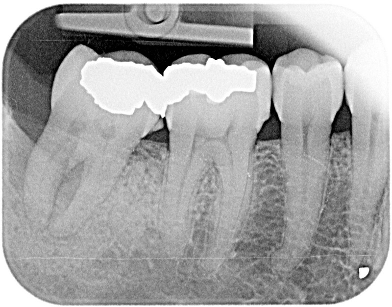
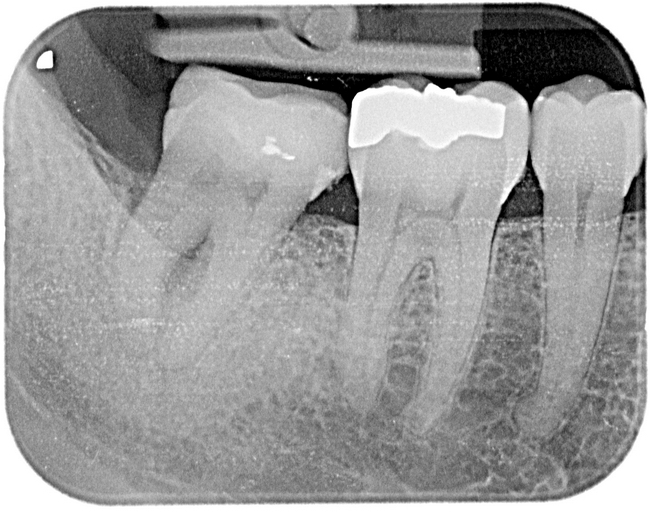
I first posted this case in Sept 2005 when this aptient visited me from out of town.This was the initial situation on tooth# 31:
size.jpg)
I took a digital pa at the time:
I treated the patient by placing a splint, using the Nd:YAG in the sulcus and adjusting the occlusion as per the advice given at the time. I saw the patient again today.
The soft tissue surrounding #31 looks good. The furcation lesion is clinically evident. The tooth went from class 3 mobility to class 1. My gut feel is that the tooth is cracked bucco-lingually. There is a visible crack, how deep it is I don’t know.
What do you see on the x-rays and where do I go from here?
Thanks very much
Etienne
etienneSpectatorSame case, tooth nuber 8. Initial situation:
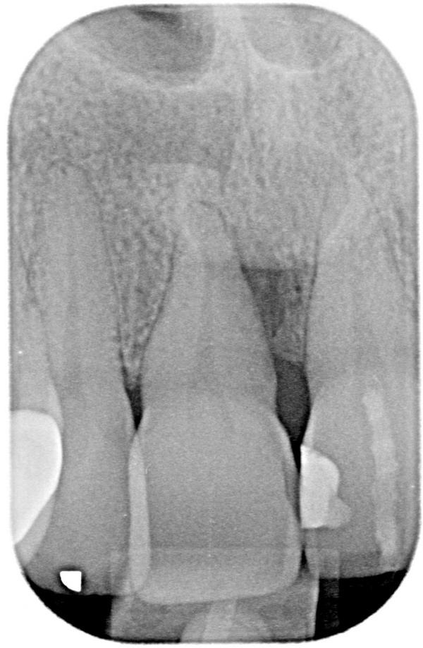
I completed endo on both #8 and #9 and splinted them.
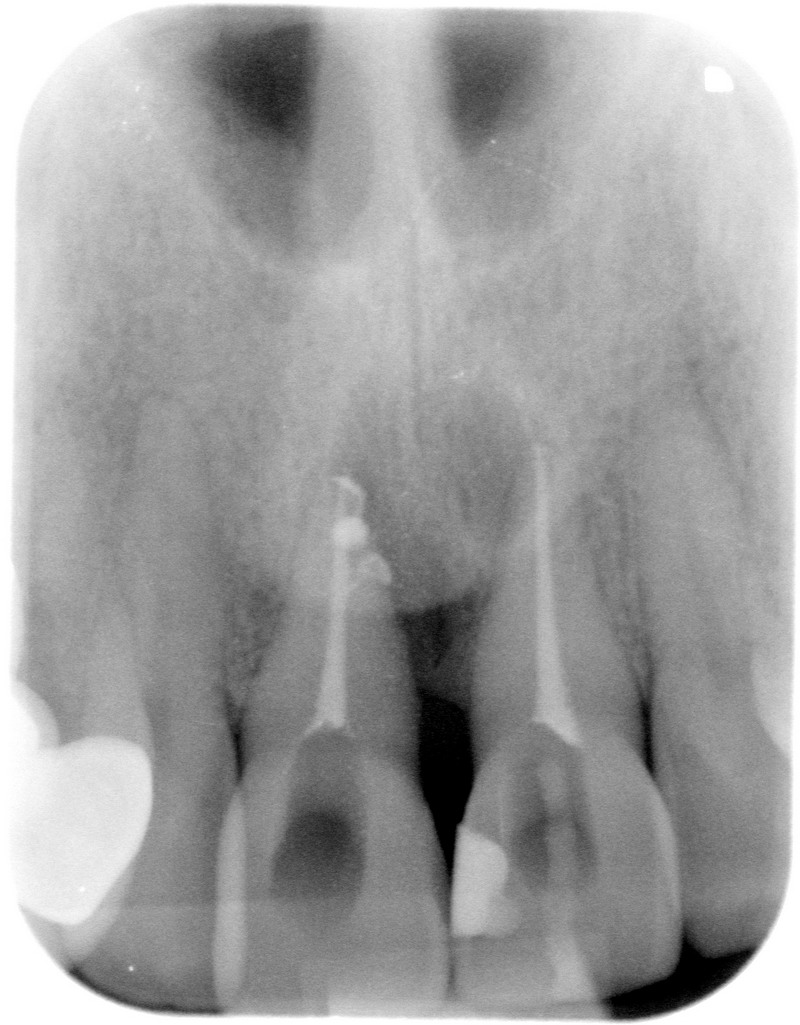
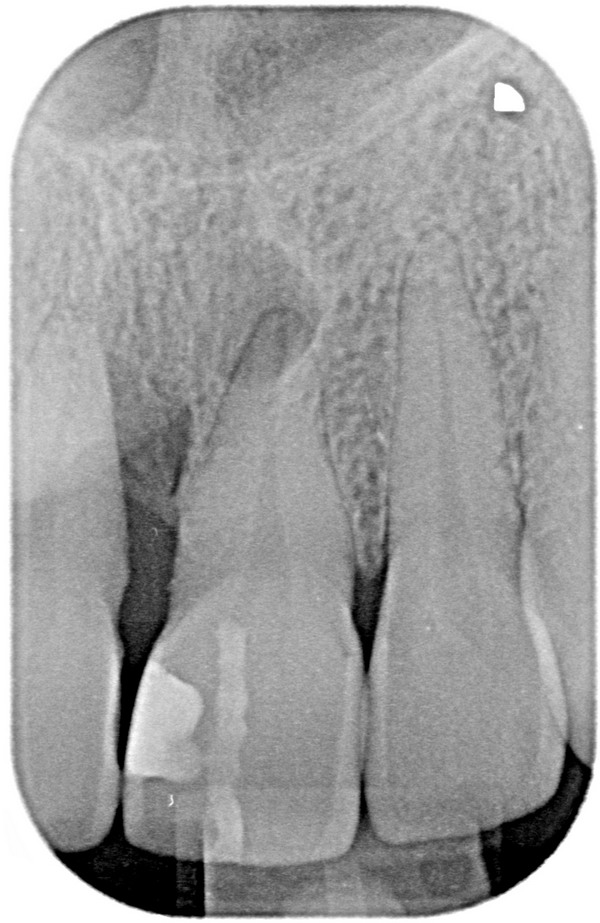
This is the current situation:
The tooth is stable because of the splint but there is still puss draining from the mesial side of #8. Vertical root fracture? There is no pain or discomfort at all.
Any advice?
Take care
Etienne
kmarshallSpectatorthis could be perio-endo. You treated the endo (great job; love the fill of the two accessory canals) now treat the perio. Hope it works out.
Keith
etienneSpectatorHi K
Thanks. My initial thought was endo-perio, which is why I did the endo. I am concerned about the fact that there is still drainage from beside the root although there seems to be more bone than at the beginning!
Take care
Etienne
etienneSpectatorHi Guys
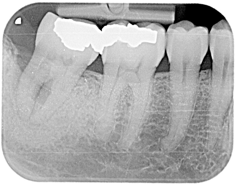
I saw this patient again in November 2006. I was disappointed in the response of the furca lesion. I re-treated the tooth in question with the laser. I also placed a temporary crown.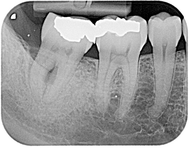
I saw the patient again this month. This is the newest x-ray.
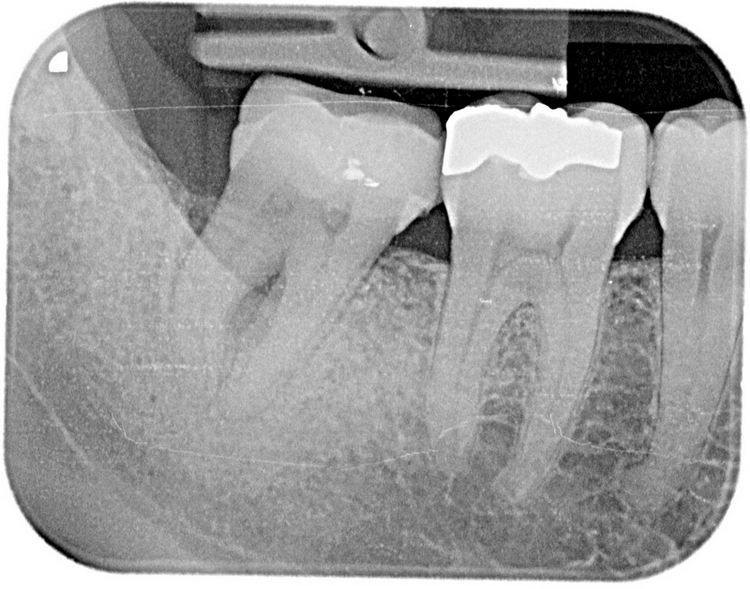
I think it is clear that the distal defect is healing. What is happening to the furca lesion? Healing? Clinically it looks good. No more drainage from the lesion. I did not want to probe yet.
Any thoughts?
Take care
Etienne(Edited by etienne at 2:23 pm on Jan. 21, 2007)
czeqm8SpectatorLook at the amount of bone in the furcation from initial to now. It is worse. At this stage, I would extract.
tschoenSpectatorI would not put any $ into this tooth unless patient insisted. If no infection is present and patient has no symptoms, what is the harm of leaving it as it is Matt?
Prognosis is certainly poor, but it is a second molar so when/if you end up removing it it is not the end of the world. If the patient really wants a tooth there, then extract and graft bone, otherwise, no news is good news.
Kenneth LukSpectatorThis can also a endo problem because there can be communication of pulpal involvement at furcation area. I’ll test for vitality. If decide to extract, open up the pulp chamber and see if the pulp is already necrotic. If so, endo first.
Ken
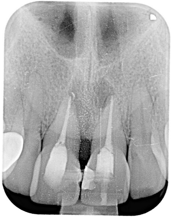
etienneSpectatorHi Guys
Further report on this case. Both the max anteriors as well as the #31 shows pocket depth reduction, lack of mobility as well as abscence of drainage from the sulcus.
At this stage I would see the treatment as being succesfull. Patient is really happy.Copy of newest x-ray:
Any thoughts?
Take care
Etienne
DinoDMDSpectatorBone appears to be filling in quite nicely Etienne.
Great job!Dino
etienneSpectatorThanks Dino
It has been a slow process but hopefully it will continue filling in, especially the furcation area. I’ll keep adding to the case pics as I monitor the patient.
Take care
Etienne -
AuthorPosts