Forums › Nd:YAG lasers › General Nd:YAG Forum › Pulsed Nd:YAG for Dentin & Decay Removal
- This topic is empty.
-
AuthorPosts
-
Robert GreggParticipantThanks to Ron and Glenn, I’m making my first attempt at taking, editing, and posting my first digital photos from my recently acquired Nikon 4500.
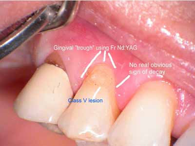
Used 4.0 Watts and 20 Hz, 100 usec PD to trough the gingiva from distal to mesial and accross the facial to expose the margin of the tooth, obtain hemostasis, and control the egress of gingival sulcular fluides.
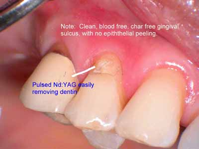
Distal area has been “prepped” using the Fr Nd:YAG at 100 usec, 3.00 watts, 300 millijoules per pulse (mj/p) and 10 Hz. Lasing was done DRY
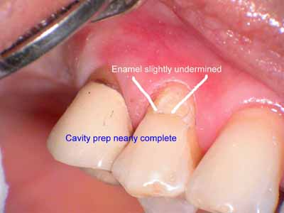
Continue removing healthy dentin, using occasional water spray from the 3-way air water syringe to remove any grey carbon (from plasma) or char that forms on the tooth surface
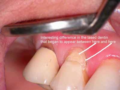
The selective nature of pulsed Nd:YAG–that “sees” different tissues with different tissue constituents, differently–allows for the tissue to ablate and appear different (Did you follow my thinking on that?) So now we have a different situation than I thought I had at first–which was to do a simple Class V. Now, because of the detection of decay in the dentin due to the pulsed 1064nm wavelength, I have a different scenario AND a different discussion with my patient.
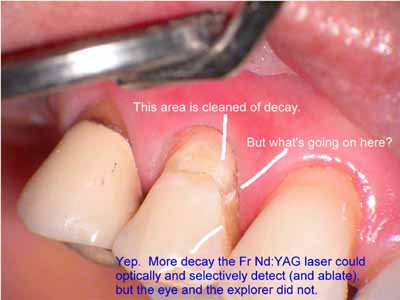
Next to last image of my prep before bond and flowable composite with fluoride. Patient informed that a full coverage crown wil be needed if the dentin around the gingiva does not remineralize. Put patient on Prevident 2x’s per day.
Bob
ASISpectatorHi Bob,
Under what magnification are these photos taken? Nice documentation. Would the decay that is not evident be detected by caries indicator or the Diagnodent?
I have also made the plunge into the scope world with a 4-step Global.
What do you think of the colour of the photos? A little faint?
What do you think? Glenn.
Regards,
Andrew
Glenn van AsSpectatorHi Bob………nicely done, they look good , a little washed out perhaps but all in all pretty darn good.
Bob, did you have it on aperture mode for the pics. just wondering and by the way…..NICE CASE.
Grin
I am getting worried that pretty soon everyone will be shooting scope photos……..
‘
FIrst Bob then Andrew……….OH MARK OH MARK……where are you MAAAAAAAAAAAARRRRRRRKKKKK.Glenn
Robert Gregg DDSSpectatorHi Andrew,
Yeah, they do look washed out. I’m playing with the settings and the image size still. With lots of help from Ron S and Glenn.
Here’s another sizing so that you can see closer up–the color and wash-out is still something to work on:
http://www.rwebstudio.com/cgi-bin/ikonboard/topic.cgi?forum=25&topic=76
Yeah, maybe caries detector or Dianodent would have worked. But as you look at the first image, there was no hint of decay to me initially. The patient will return in a couple of weeks and I’ll put some indicator on and see what it looks like. I don’t have a Diagnodent.
I used .66 magnification with I think is 4x. Glenn?
What’s the translation for those numbers on the 5 Step anyway (and why in the heck doesn’t Global just replace or add the mag number on the dial?!)
Thanks Glenn for the very GENEROUS remarks. I’m getting better thanks to you, Ron and Ralph Klink. I’ll check the aperture mode. The pics have some noise (ISO now at 200) and color issues still.
Don’t worry TOO much Glenn, in takes a while to get used to the scope in general before positioning the camera in addition to that.;) I’ve had 5 years or so with the scope so I know how to position the scope, steer it, fine focus it, hold it steady, “nose it” around, then take the picture.
But this IS fun:cheesy: , and outstanding for sharing clinical concepts, technique and technlogy of lasers and scopes. And because it is so easy to take the shot through the scope while working–instead of stopping to get the 35mm–there will be fewer missed opportunities to share clinical surprises like the one exampled here……
Thanks for the feedback, I appreciate it.
Bob
PS You know, I guess I should add and point out that the dentin looks white because it has been etched with the Nd:YAG. It melts and resolidifies dentin 60-100 microns deep and chemically as well as mechanically alters the dentin by removing the inorganic components and increasing the amount of mineral content.
(Edited by Robert Gregg DDS at 10:09 am on May 15, 2003)
Glenn van AsSpectatorHi Bob: depending on the length of your objective lens (200mm or 250 mm) and your eyepieces (most are 10X, but can be 8X or 12.5X mag) the .5 turret is 4x mag and the .66 is about 6X mag.
The magnification changes alot ………..depending on your objective and eyepieces.
For instance if you have a 200mm objective lens and 10X eyepieces than each number is multiplied by 8.
If you instead have a 250 mm objective lens you multiply each number by only 6.3. Its quite a difference for instance if you take the 1.25X tur ret spot and multiply by 8 you have 10X mag but if you only multiply by 6.3 then you have around 8x mag.
The 8X eyepieces will lower the mag again and the 12.5 X mag raises them.
It really is amazing to not only see the cases through the scope but to photograph them. Look how power Marks recent photos of how to prep a Class 1 with the laser are because of the closeup.
I am sure that with time he will get a scope and a camera, because the ease with taking photos right through the scope is unparalleled.
Cameras and scopes take a while to get the sweet spot.
I will ask Eric if he has any PDF files for using the Xmount adapter for the scope with your Nikon 4500.
Glenn
Robert GreggParticipantGlenn,
Ralph has me set at S Shutter 1/125
What up with that?
Bob
pcpackerSpectatorI am learning a lot about lasers, as I read this forum. I didn’t even know that the periolase could cut hard tissue. How would it perform around a crown margin with recurrent decay (like the second molar in the above photos)?
Robert Gregg DDSSpectatorHowdy pcpacker,
Yes, for over 13 years Nd:YAG laser that are pulsed in the millionths of a second (microseconds) like pulsed erbium YAG and YSGG dedicated hard tissue lasers have been well known to cut dentin, diseased enamel, caries. The pulsed Nd:YAGs do NOT cut healthy enamel, and that’s what a lot of people think about when they are interested in a “hard” tissue laser.
And since few companies exist anymore that manufacture and sell pulsed Nd:YAGs, most are unaware of this capability.
To answer your question, it will do just fine around a crown margin in removing decay. The quartz fibers are pretty rugged, so you don’t need to worry about that. Since all lasers are “end” cutting, all lasers have difficulty getting under a crown margin. But when I need to treat under a crown margin, I just cut it away with a bur or diamond. So, sure, it will do just fine. This particular crown has a failing RCT, so I need to cut it off.
If I have time, I could remove the decay with the PerioLase, take photos, remove the crown and see how I did, then come back and post it here. I think we are about ready to take this tooth on–she had advanced perio and many failing RCTs that were worse).
The PerioLase was design to have maximum periodontal disease treatment parameters, AND hard tissue parameters.
I use the PerioLase to:
1. Locate and open calcified canals in endo.
2. Detect and remove enamel decay in pits & fissures.
3. Detect and remove dentin decay.
4. Melt and resolidify dentin around cavity prep margins to increase caries resistance upwards of 40%.
5. Etch dentin to remove collagen, increase mineral matrix, and bond to mineral.
6. Etch dentin to reduce post resin sensitivity.
7. Etch dentin to increase bond strength around 70%.These are just a few hard issue uses.
We named the PerioLase and develped its parameters for the disease that we struggle so much in dentistry to resolve–and because no other laser wavelength has the selectivity to address the major challenges in pero disease treatment–not because it can’t do other things as well like hard tissue.
I congratulate you on your investigations into lasers to get beyond hype and really learn what various lasers can do.
Thanks for the question.
Bob
BNelsonSpectatorHi Bob
Just finished two LPTs today and agree that the Periolase is much more useful than just perio tx. The selective removal of enamel caries always amazes patients, and the ability to see into the canal to chase calcified canals is really great- I can work with my lopes ( no microscope, yet) and see whereas with a handpiece, it’s always in the way. After talking to Glenn at AMED I have to get a scope soon. Those pictures are just so cool and I feel like I can’t see much anymore.
Glenn van AsSpectatorBruce: It was an absolute pleasure for me to talk to you at the meeting. I think it is awesome that someone takes the time to seriously look at the technology. I can tell you I have TONS of dagger marks from DT in my back over the scope issue. People feel intimidated, worried that they have to move to a scope, defensive, and defiant. I only want them to have an open mind. I think it is wondeful to use the scope for all dentistry, but I gotta tell you that many times each and every day now I think……..hmm……..is it overkill. I wonder if guys like Rod from DT and Mauty are right.
THen it occurs each and every day that I work…….I wouldnt have seen that if I hadnt been using the scope. Today it was a lower first molar heavily decayed that I split in half with the erbium Yag and removed bone around each root and then elevated both roots out one at a time. I could tell from the scope when I had a good purchase point and when the root started to move not by feel………I COULD SEE IT MICROMOVING.
Its so hard to explain to people that sometimes I really feel like just saying its not worth it……….then people like you post things like this and I know its all worth it. I know I have found my mission in life…….scopes are fun, make my dentistry exciting and improve the precision of things. When people like you Bruce approach it with an open mind, you make me realize that it is all worth the daggers.
Thanks Bruce, its with a huge heartfelt response that I want you to know how I appreciate that simple post……..
It made my whole day.
Glenn
vinceSpectatorHi Guys,
I demo’d a scope in my office for a long time. Thanks Global and Steve. Once you try it….it’s REALLY HARD not to use it (I have DFV 3.5x loupes). What you can see is astounding. I have ordered my scope and am waiting for it to arrive now. The most recent endo I did felt like I was going about it ‘blind’ after using the scope for it. Lasers, microdentistry, and scopes are a wicked combo. Then you have the video/photo capabilities as well. If you haven’t tried it, do so. It won’t cost you anything.
Regards.
Glenn van AsSpectatorThanks Vince: it is gratifying to see others also following the pathway of scopes and lasers. In my opinion they are a natural combo and the patients are the big winners in my practice.
Documentation is a breeze and video documentation is coming along and will revolutionize the way we document cases when some of the compression systems that I here about for video become commonplace.
Photos will be out and videos will be in………WONT THAT BE COOL HUH.
HDTV is coming and it will allow you to grab images and video from the same source……..wow that will be awesome.
Its a great ol time to be a dentist…….lasers , scopes , and all sorts of digital cameras…….man you can play with more tools than you can shake a stick at!!
Cya and Vince………..thanks
Glenn
ASISpectatorHi Vince & Glenn,
Congrats, Vince.
Hey, Glenn, another enlightened dentist converted.
How wonderful! One can tell who is willing to step up and make that leap to practise a higher level of dentistry from one’s own bench mark….
Andrew
Glenn van AsSpectatorIt comes in small numbers Andrew……a few people see the light ( laser light that is!!) and they realize that magnifcation really is important.
Mark Colonna is next on my list and David Kimmel is trying to hide, whimpering in the background behind Allen and Albert……….
David IT IS FUTILE TO RESIST, come Join the dark side, those using scopes.
THERE ARE ENOUGH DAGGERS FOR ALL!!
Mauty and Rod from DT cant only lambaste me!!
Grin
Glenn
-
AuthorPosts
