
Forums › Diode Lasers › General Diode Forum › Treating Peri-implantitis with 980
- This topic is empty.
-
AuthorPosts
-
Kenneth LukSpectatorBob,
I’m confused. Please show me the ‘light’ end of the tunnel and forgive me for asking an ignorant question.NIR is transparent through water and is not absorbed in tissue. When laser hit tissue, the interaction is ‘Heat altered proteins’. Does this not contradict?
Ken
(Edited by Kenneth Luk at 12:49 pm on June 18, 2003)
Robert Gregg DDSSpectatorSorry Ken,
Typo.
I didn’t mean in “tissue”. I meant in “water”.
So you are correct. My statement was a contradiction.
Thanks for pointing that out.
I will edit to correct.
Bob
Kenneth LukSpectatorThanks Bob,
The paper I quoted was only trying to point out that there is no damage on titanium surface making 980 pretty safe for implant recovery and peri-implantitis.Bob, the LPT case you posted on soft tissue section was very interesting. Please keep us posted on this case.
I’m sure you’ve heard of perio2000 by Diamond General Development Corp. http://www.perio2000.com
Do you think it may show us the differences in bacterial activities in the pockets before and after LPT, 6 mths post-op?
What’s your view on perioscope?(Edited by Kenneth Luk at 11:38 am on June 19, 2003)
(Edited by Kenneth Luk at 11:41 am on June 19, 2003)
Kenneth LukSpectatorBob,
I need your “light ” to show me the way again! Please bare with more basic questions!
In your post on perio treatment with waterlase, you mentioned that:
NIR transmitt through water in tissues with NO attenuation of the beam intensity until the beam hits a pigment the wavelength is highly absorbed into– like black pigmented anaerobes embedded out into the tissues.
In this post you mentioned when laser hits tissue :Heat altered protein.
Does that mean healthy tissue is also affected as well as bugs? Lasers penetrate through water to different depths. How deep do these bugs infiltrate into gum tissues?
Granulation tissue has a higher water content than healthy tissue. Isn’t the higher water content in infected tissue more prone to vaporisation ?
Thanks
Ken
ASISpectatorHi Ken,
When using your irrigation handpiece, are you using it with the ultradent canula tip?
Andrew
Kenneth LukSpectatorhi Andrew,
Yes, I use the Ultradent canula tip.
Ken
ASISpectatorHi Ken,
Do you not find that the water tends to have trouble flowing out the tip of the canula? Perhaps I haven’t adjusted the flow correctly?
Andrew
Kenneth LukSpectatorHi Andrew,
Yes, there were problems.
After speaking to Joe Mooney, the engineer, here’s what I’m doing now:
If you use the water from the unit , the flow is too much and there’s leakage at the middle and the end of the handpiece. To avoid leakage, give it some torque when tightening the handpiece.Also not keen on using water from the unit.I would prefer to use sterile saline pack for irrigation.
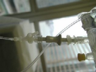
Joe made me this connection to the saline pack.The pack is tided to the arm of the ceiling light mount. The flow is controlled by a valve at either end of the tubing.
Size 20 ultradent gauge used on 360 fiber.I did 8 quadrants of laser curretage with SRP , troughing
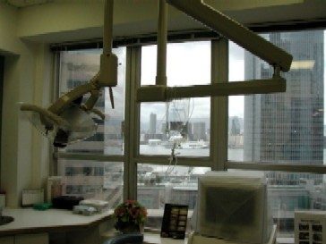
on two crown preps today. Almost finished the saline pack. Once the pack is opened, we discard it in one week BUT we always managed to finish the pack within the time.Photos attached. Thought I’d show you the harbour view of HongKong from my office.
Saline pack connection.
size 20 canula with 360 fiber.
Hope this is useful for you.
You can contact Joe at Biolitec.Ken
 3.jpg)
(Edited by Kenneth Luk at 11:37 am on June 23, 2003)
(Edited by Kenneth Luk at 11:47 am on June 23, 2003)
(Edited by Kenneth Luk at 8:41 pm on June 23, 2003)
Robert GreggParticipantHi Ken and Everyone,
Sorry for being away for so long. Busy, busy, busy with clinical practice and training, dentists, and hygienists from out of town.
Not to mention the strange, stray Canadian Canucklehead that came by for dinner….None other than Glenn van As! It was GREAT to have him visit. Glenn even offered to pay for EVERYONE’s dinner. Thanks, Glenn! What a great guy……..I’ll send you the receipt right away…..:cheesy:
Ken, you asked, “Bob, the LPT case you posted on soft tissue section was very interesting. Please keep us posted on this case.”
You bet. The patient is out of town and will be back next Wednesday. I’ll post his post op progress.
“I’m sure you’ve heard of perio2000 by Diamond General Development Corp. http://www.perio2000.com
Do you think it may show us the differences in bacterial activities in the pockets before and after LPT, 6 mths post-op?”Yes, I have heard of them and their probe. The Diamond probe detects levels of sulfide from sulfide producing (H2S) bacteria.
It might help to some degree in detecting levels of bacteria at 6 months LPT, but there are LOTS of other things to attend to that contribute to periodontal disease or health than P. gingivalis, P. intermedia, and B. forsythus.
In fact, these “opportunistic” anaerobes mentioned above are NOT the first to invade the pocket, and probably would not be present at 6 months post LPT if all is going as expected–unless they were not effectively killed in their “priviledged” sites (i.e. endothelial cells, macrophages, calculus, dentinal tubules) during LPT treatment.
So the Diamond probe might not be of much help for the gram positive bugs that first populate a developing pocket.
“What’s your view on perioscope?”
The perioscope is the first generation dental endoscope. I think the idea is great, and the first version is fine, but I think we will see better imaging and scopes in the future.
“In your post on perio treatment with waterlase, you mentioned that: NIR transmitt through water in tissues with NO attenuation of the beam intensity until the beam hits a pigment the wavelength is highly absorbed into– like black pigmented anaerobes embedded out into the tissues.” Correct
“In this post you mentioned when laser hits tissue (that it is absorbed into) :Heat altered protein.” Correct
“Does that mean healthy tissue is also affected as well as bugs?”
Correct, if it is healthy tissue that is absorbing the particular wavelength.
“Lasers penetrate through water to different depths.”
Correct. That’s the “coefficient of absorbtion in water” chart we all have seen so much of.
“How deep do these bugs infiltrate into gum tissues?”
Just like a pimple can expand into a boil, or an abcessed tooth can spread into the cavernous sinus behind the eye, the anaerobes can penetrate many centimeters into tissue.
“Granulation tissue has a higher water content than healthy tissue. Isn’t the higher water content in infected tissue more prone to vaporisation ?
Your statement and follow-on question presumes that granulation tissue is not healthy. To quote Orban’s Perio 1972, “Granulation tissue is found in healing wounds and is composed of fibroblasts and proliferating capillaries, which also give it a granular appearance. With healing, granulation tissue reforms into a normal fibrous connective tissue.”
With NIR lasers, tissue–granulation or otherwise–infiltrated with bacteria that highly absorb the laser’s wavelength would tend to vaporize more quickly, watery tissue or not!;)
Bob
ASISpectatorThanks Ken.
Nice view of the harbour.
Andrew
Kenneth LukSpectatorHi Bob,
Thanks for your comprehensive reply!Ken
dkimmelSpectatorI have a Implant patient with recurrent peri-implantitis. The implants are over 11 years old. The osseous contours around three of the implants are not ideal.
[img]https://www.laserdentistryforum.com/attachments/upload/IMG3efb6dd3.JPG[/img]
The bone levels have not changed over the 3 years I have seen this patient. There is subcalculus present and the crown and bridge is not ideal. All of which a feel contribute to the problems of this case.
How would you intergate the use of the Lasersmile in the treatment of this site?
David -
AuthorPosts