
Forum Replies Created
-
AuthorPosts
-
Glenn van AsSpectatorHi Tom …….have a safe trip to Florida and continued success man with all you are doing.
National Dental Network is a good organization and really does a nice job on the stuff I have seen.
I am up for the improvements in the guideline. Heck if I use 10% of what I have learned from all the gurus here (even the Biolase guys – joke joke) then it will be a good addition for new users.
Great to see you online here……..drop by more often. Almost 400 people here now…..its a big site.
Cya
Glenn
dkimmelSpectatorGlenn,
I used no anesthetic. This was with water and defoused. There was no bleeding in this mode and the tissue came off in thin layers. Around #7 I had wanted to thin the paplallia and this worked great. Nopost op discomfort which I would have expected if the watts were too high. There was one area on the mesial of #7 that had a concavity. In this area I dropped back to .25w with 11% a, you will see this as the red area in the photo. Just too tight a space to use the high watts.
#7 is a peg lateral. The kid is 18 y/o. She has had some pretty bad ortho. Not only is the roots on #7 resorbed but #6,8,9 & 11. She may loose all of them. To top it off the plan was to place an implant in #10 spot ,post ortho #9 roots were tilted in the postion where the implant was to go. She had to go back to ortho. I did not get involved in this case until the patient walked in wanting the implants after the first ortho.
Sticktech– cool stuff. Like ribbion but much easier to work with. I don’t think it is on the market yet. Let me know if you want samples. I’ll give you the contact.
The ovate pontic. Yes and no. Sort of. I did not have much depth to work with .The photo is not the best. I have about a 2mm depth after preparing the site and 2mm to bone. I was trying to push the papallia forward. Still got to work on that as it is back too far at this point.
David
whitertthSpectatorDavid
Great stuff…Nice use of composite, great shade match….. very nice result….What is stichtek and where can u get it? Are most people using the waterlase/hoya units on soft tissue dry without water and without anaesthsia? Nice work again!!
whitertthSpectatorGreat stuff glenn!!!! If u remember i did something similar a while back with an exposure…cauterized ( mine with the waterlase) and etched and bonded nad we are at 6 months so far…so who knows with this stuff…maybe we r onto something….As far as MTA I have never used it although have heard about it….can u enlighten me on when u use it etc…….Thanks again
AlbodmdSpectatorThis isn’t specifically laser literature, but it’s some literature comaparing MTA and Portland Cement (which is a lot cheaper) that I copied from DentalTown.
Regards,
Al BMTA vs. Portland cement–the literature speaks:
1:Aust Endod J. 2003 Apr;29(1):43-4.
A comparative analysis of Mineral Trioxide Aggregate and Portland cement.
Funteas UR, Wallace JA, Fochtman EW.
The purpose of this study was to compare the composition of Portland cement and
Mineral Trioxide Aggregate (MTA). Samples of MTA and Portland cement were
analysed for fifteen different elements by inductively coupled plasma emission
spectrometry (ICP-ES). Comparative analysis revealed there was significant
similarity except there was no detectable quantity of Bismuth in Portland
cement. Quantitative results are given in both parts per million (p.p.m.) and
wt%. It was concluded that there is no significant difference between the 14
different elements in both Portland cement and MTA.2: Oral Surg Oral Med Oral Pathol Oral Radiol Endod. 2003 Apr;95(4):483-9.
Cell and tissue reactions to mineral trioxide aggregate and portland cement.
Saidon J, He J, Zhu Q, Safavi K, Spangberg LS.Objective. Mineral trioxide aggregate (MTA) is being widely used for root-end
fillings, pulp capping, perforation repairs, and other endodontic procedures.
MTA and Portland cement (PC) have many similar physical, chemical, and biologic
properties. PC cement has shown promising potential as an endodontic material in
several studies in vitro and in vivo. The purpose of this study was to compare
the cytotoxic effect in vitro and the tissue reaction of MTA and Portland cement
in bone implantation in the mandibles of guinea pigs. Study Design. Millipore
culture plate inserts with freshly mixed or set material were placed into the
culture plates with already attached L929 cells. After an incubation period of 3
days, the cell morphology and cell counts were studied. Adult male guinea pigs
under strict asepsis were anesthetized, during which a submandibular incision
was made to expose the symphysis of the mandible. Bilateral bone cavities were
prepared and Teflon applicators with freshly mixed materials were inserted into
the bone cavities. Each animal received 2 implants, one filled with ProRoot and
1 with PC. The animals were killed after 2 or 12 weeks, and the tissues were
processed for histologic evaluation by means of light microscopy. Results. There
was no difference in cell reactions in vitro. Bone healing and minimal
inflammatory response adjacent to ProRoot and PC implants were observed in both
experimental periods, suggesting that both materials are well tolerated.
Conclusions. MTA and PC show comparative biocompatibility when evaluated in
vitro and in vivo. The results suggest that PC has the potential to be used as a
less expensive root-end filling material.3: Braz Dent J. 2001;12(2):109-13.
Healing process of dog dental pulp after pulpotomy and pulp covering with
mineral trioxide aggregate or Portland cement.
Holland R, de Souza V, Murata SS, Nery MJ, Bernabe PF, Otoboni Filho JA, Dezan
Junior E.
Considering several reports about the similarity between the chemical
compositions of the mineral trioxide aggregate (MTA) and Portland cement (PC),
the subject of this investigation was to analyze the behavior of dog dental pulp
after pulpotomy and direct pulp protection with these materials. After
pulpotomy, the pulp stumps of 26 roots of dog teeth were protected with MTA or
PC. Sixty days after treatment, the animal was sacrificed and the specimens
removed and prepared for histomorphological analysis. There was a complete
tubular hard tissue bridge in almost all specimens. In conclusion, MTA and PC
show similar comparative results when used in direct pulp protection after
pulpotomy.
Glenn van AsSpectatorThanks David, and yes Ron I am using the Hoya without water and without anesthesia.
I typically am in contact or slightly away at around 30 Hz and 30-50 mj which is .9-1.5 w
If the patient feels it even with air blowing then sometimes I drop to around .6 w with 20 Hz and 30 mj.
Nice work David and thanks for the answers. Looking forward to seeing Stick tech.
You didnt prepare the adjacent teeth did you?
Glenn
Glenn van AsSpectatorExcellent stuff Albert, and I mean it.
I hear that the MTA is sterilized and that is it and otherwise it is PC.
I havent used the white stuff yet , apparently it handles differently and also may not set as well.
I only use a small amount out of the 100 dollar package, per patient.
It is tough to handle (like sand really) and I try to mix it dry.
Joe Dovgan makes a set of three pluggers called the Dovgan pluggers which are nice carriers and in different sizes. I will look to see who supplies them.
MTA I use mainly in perfs, or in large open canals which dont calcify over with CaOH. It also has been shown by many to be effective in pulp caps as it is biocompatible.
Some use it in apicos as well as the root end filling material.
You can get it from Dentstply and it is expensive. The powder you just mix with water , and in addition it is a little tough to handle the first couple of times you use it.
Cya and thanks
Glenn
dkimmelSpectatorHere is a link to another case using the StickTeck
http://www.dentaltown.com/gold….D40B3EC
Here is the companies web site.
http://www.sticktech.com
The person I’ve talked with is Dr.Elja Korhonen
his e-mail is elja.korhonen@sticktech.comI did prep the linguals with a small groove like the above case. I was trying not to post tooo many pictures. Ron still has to go back and clean up my posts
for me. I’ll get there.
Glenn the more I most the more I understand the effort it takes to post all the photos you do. Thanks again.
David
ASISpectatorHi Glenn & Bob & All you other Keeners
In use of the diode to pulp cap, are you in contact or near contact to coagulate the exposure?
Thanks again.
Andrew
2thlaserSpectatorGlenn, I have been using Messing Guns for delivery of the MTA. They have very small orifi for delivery, mainly for use in apico’s, but I have found it very useful in placing MTA in cases like you have above. Great stuff.
Thanks for sharing this stuff. I need a scope, just need the $ for it first (wife thing!!).
Mark
Kenneth LukSpectatorHi Glenn,
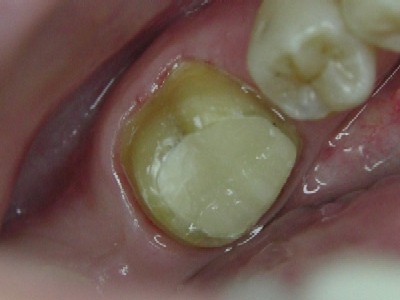
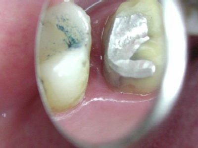
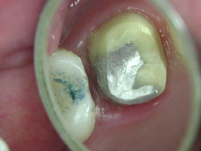
I did a similar case recently.
Patient ( Female, 20 )attended with fractured mesial lingual cusp.Carious exposure after amalgam removal.
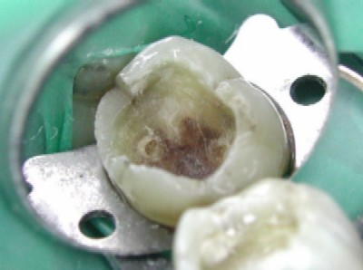
Coagulated exposure with 980
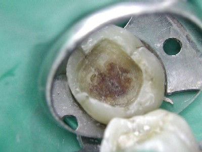
Placed MTA on the area.

I have an endodontist at my office and he was advicing on placing a very thin layer of calcium hydroxide over the MTA (to be safe to ensure MTA sets hard). He was saying that sometimes the MTA does not set rock solid. He tend to check the MTA on a second visit at times when the exposure is large.
(He was saying that the MTA does not set well on acidic condition. Also MTA requires some moisture to set.)
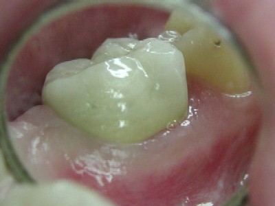
No photo showing calcium hydroxide placement.Photac fill ( compomer) lined the cavity
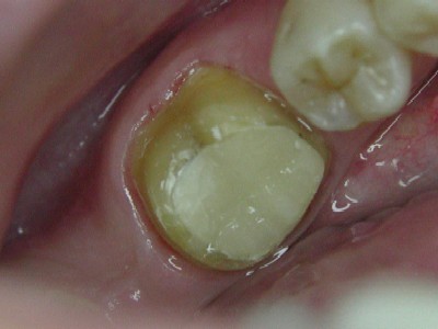
To avoid futher tooth fracture, Crown prepped and troughed with 980 ( 1w cw )
Impression
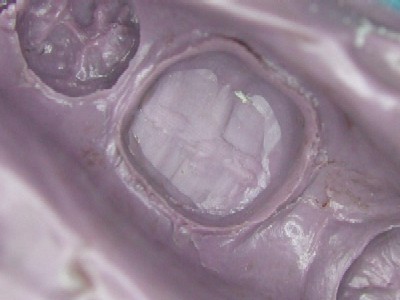
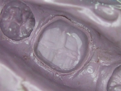
Troughed margin 1 wk post-op
Crown fitted
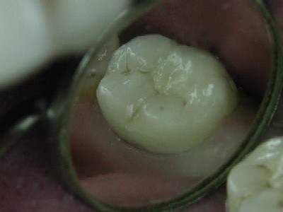
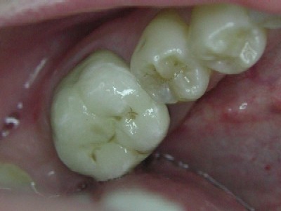

She experienced no symptoms during the week.
She was also made aware of possibility of future RCT.Ken
(Edited by Kenneth Luk at 4:51 pm on July 16, 2003)
(Edited by Kenneth Luk at 4:53 pm on July 16, 2003)
(Edited by Kenneth Luk at 5:00 pm on July 16, 2003)
AlbodmdSpectatorI was looking at articles relating on how lasers effect bond strength and I found these. I had always thought that lasers increase bond strength, but these articles seem to state otherwise. Anyone’s thoughts?
: J Clin Laser Med Surg. 2003 Apr;21(2):105-8. Links
Influence of the frequency of Er:YAG laser on the bond strength of dental enamel.
Goncalves M, Corona SA, Pecora JD, Dibb RG.
School of Dentistry of Ribeirao Preto, University of Sao Paulo, Sao Paulo, Brazil.
OBJECTIVE: The present study had the aim of evaluating the influence of different frequencies of the Er:YAG laser on adhesive resistance of enamel and one restorative system. Background Data: There have been no reports of studies assessing the influence of the pulse frequency variation of the Er:YAG laser on adhesive resistance of the enamel/resin interface. MATERIALS AND METHODS: Fifty surfaces of enamel from extracted human third molars were planed and divided into five groups at random. Enamel surface treatment was realized by the Er:YAG laser at 80-mJ power and 1-, 2-, 3-, and 4-Hz pulse frequencies, followed by etching. For the control group, only acid conditioning with 37% phosphoric acid for 15 sec was used. The Single Bond/Filtek Z250 system was chosen for the fabrication of the specimens, which were stored in 100% relative humidity for 24 h, at 37 degrees C. The specimens were submitted to tensile resistance tests using a Universal Testing Machine (50 Kgf and 0.5 mm/min). RESULTS: The mean values in MPa were 1 Hz, 25.58 (+/-6.16); 2 Hz, 25.58 (+/-3.79); 3 Hz, 21.34 (+/-3.78); 4 Hz, 21.17 (+/-3.13); and phosphoric acid only, 22.44 (+/-7.0). Data were submitted to statistical analysis using ANOVA, and there was no significant difference in tensile resistance between the studied groups.
CONCLUSION: The results suggest that the Er:YAG laser, with 80-mJ power associated with acid conditioning at 1-, 2-, 3-, and 4-Hz frequencies, did not present significant improvement in tensile bonding of enamel, as compared to acid conditioning only.J Dent. 2003 Feb;31(2):127-135. Related Articles, Links
SEM evaluation of the interaction pattern between dentin and resin after cavity preparation using ER:YAG laser.
Schein MT, Bocangel JS, Nogueira GE, Schein PA.
Abdon Batista 121, sala 904 Centro, 89201-010, SC, Joinville, Brazil
Objective. The aim of this study was to describe the interaction pattern formed between dentin and resin on cavities prepared with an erbium laser (Er:YAG). The morphological aspect of the irradiated dentin after acid etching was also observed.Methods. Ten dentin disks were obtained from fresh extracted third molars. Each disk received two cavities, one prepared with a conventional high-speed drill, while the other cavity was obtained by the use of an Er:YAG laser (KaVo KEY Laser, KaVo Co.). The laser treatment was performed with 250mJ/pulse, 4Hz, non contact mode, focused beam, and a fine water mist was used. Five disks were prepared for morphological analysis of the acid etched dentin. The other five disks had their cavities restored with Single Bond (3M) followed by Z100 resin (3M). The specimens were observed under scanning electron microscopy after dentin-resin interface demineralization and deproteinization.
Results and conclusions. It was observed that the morphological characteristics of the acid-etched irradiated dentin were not favorable to the diffussion of monomers through the collagen network. The dentin-resin interfacial aspect of irradiated dentin, after acid etching, showed thin tags and scarce hybridization zones, which agreed with the morphology of the irradiated and acid-etched dentin substrate observed.Eur J Oral Sci. 2002 Aug;110(4):322-9. Related Articles, Links
Micro-tensile bond strength of two adhesives to Erbium:YAG-lased vs. bur-cut enamel and dentin.
De Munck J, Van Meerbeek B, Yudhira R, Lambrechts P, Vanherle G.
Department of Conservative Dentistry, School of Dentistry, Oral Pathology and Maxillo-Facial Surgery, Catholic University of Leuven, Belgium.
The purpose of the study was to assess the hypotheses that laser irradiation is equally effective for bonding as traditional acid-etch procedures, and that tooth substrate prepared either by Erbium:YAG laser or diamond bur is equally receptive to adhesive procedures. Buccal/oral enamel and mid-coronal dentin were laser-irradiated using an Erbium:YAG laser. A total-etch adhesive (OptiBond FL) applied with and without prior acid-etching and a self-etch adhesive (Clearfil SE Bond) were employed to bond the composite. The micro-tensile bond strength (microTBS) was determined after 24 h of storage in water. Failure patterns were analysed using a stereo-microscope, and samples were processed for Field-emission Scanning Electron Microscopy (Fe-SEM) evaluation. Unbonded, lased enamel and dentin surfaces were evaluated using Fe-SEM as well. The total-etch adhesive bonded significantly less effectively to lased than to bur-cut enamel/dentin. Laser ‘conditioning’ was clearly less effective than acid-etching. Moreover, acid etching lased enamel and dentin significantly improved the microTBS of OptiBond FL. The self-etch adhesive performed equally to lased as to bur-cut enamel, but significantly less effectively to lased than to bur-cut dentin.
It is concluded that cavities prepared by laser appear less receptive to adhesive procedures than conventional bur-cut cavities.
Kenneth LukSpectatorHi Glenn,
I’ve been troughing with gated pulse at 4-6 w with irrigation with 980.
I tried one recently with 1w cw.
Here’re the post-op views of the tissues.Gated pulse 4 w with irrigation
Tissue was inflammed pre-op
1w cw without irrigation
With higher power and irrigation, there’s no smell of charring on the fiber.
With 1w cw , I moved the fiber pretty quickly but still get a bit of burning smell. I suppose the suction took care with most of it.Wish I have a mic to capture better pictures!
Ken
Kenneth LukSpectatorGlenn,
Participations on your proposed hands-on will be overwhelming.
You may have to repeat the hands-on throughout the ALD meeting.
Reserve me a place please!!
Ken
ASISpectatorHi Ken,
Nice treatment. What setting did you use to coagulate and was it noncontact or contact?
Andrew
-
AuthorPosts